LLLT Device How It Works: The Complete Science Behind Cold Laser Therapy
Summarized from peer-reviewed research indexed in PubMed. See citations below.
Get our free Cold laser therapy research guide
Evidence-based insights delivered to your inbox
Understanding how LLLT devices work starts with photon-tissue interaction. Research shows that specific wavelengths of light can increase cellular energy production by 150-200% when properly absorbed by mitochondrial chromophores. The Cold Laser Human/Vet Device with LED Display 2x808nm +12X650nm ($129) delivers both 808nm near-infrared and 650nm red wavelengths at 150mW total power, providing the optimal spectral range for cytochrome c oxidase activation and tissue penetration. This dual-wavelength approach activates mitochondrial electron transport chains while stimulating collagen synthesis, supported by studies showing 40-60% reductions in inflammatory markers within 48 hours. For budget-conscious users, the Cold Laser Therapy Device 4×808nm + 14×650nm with LCD Display ($79) provides similar wavelength coverage with 140mW total power across 18 diodes. Here’s what the published research shows about the cellular mechanisms that make LLLT effective.
Disclosure: We may earn a commission from links on this page at no extra cost to you. Affiliate relationships never influence our ratings. Full policy →
How Does Light Energy Interact with Biological Tissue?
Light interaction with tissue begins with photon absorption by chromophores, which are molecules that absorb specific wavelengths. When photons in the 600-900nm range enter tissue, they encounter water, melanin, hemoglobin, and cytochrome c oxidase. Each chromophore has distinct absorption spectra that determine which wavelengths get absorbed versus scattered or transmitted.
Cytochrome c oxidase, the terminal enzyme in the mitochondrial electron transport chain, absorbs wavelengths between 600-900nm with peak absorption around 620nm, 680nm, 760nm, and 820nm. A 2017 study published in Photochemistry and Photobiology measured these absorption peaks in isolated mitochondria, demonstrating that 808nm light produces the strongest increase in oxygen consumption rates. This wavelength specificity is why choosing the right cold laser therapy device matters — devices must emit within this absorption window to activate the primary photoacceptor.
The depth of light penetration depends on wavelength and tissue composition. Red light at 650nm penetrates approximately 8-10mm through skin and subcutaneous tissue before intensity drops to 37% of surface irradiance. Near-infrared light at 808nm penetrates 30-40mm, reaching deeper structures like muscle tissue, tendons, and superficial joints.
Scattering reduces light intensity as photons bounce between cellular structures, collagen fibers, and extracellular matrix components. This scattering follows Mie theory for particles similar in size to wavelength, creating a diffuse light field within tissue. Research in Lasers in Surgery and Medicine used Monte Carlo modeling to show that 808nm light scatters less than 650nm, explaining superior deep-tissue penetration.
Absorption by water increases above 900nm, which is why wavelengths like 980nm and 1064nm generate more heat and less photobiomodulation. The “optical window” between 600-900nm minimizes water absorption while maximizing chromophore activation, creating therapeutic effects without thermal damage.
Power density (irradiance) at the skin surface determines how much energy reaches target tissues. A device delivering 100 mW/cm² at the surface provides approximately 20-30 mW/cm² at 20mm depth with 808nm light, depending on tissue composition. This depth-dependent reduction means application time must increase for deeper structures.
Coherence, a property unique to laser light, maintains spatial and temporal alignment of photons. Studies comparing coherent laser to incoherent LED light at identical wavelengths and power densities show mixed results, with some research finding no significant difference in cellular outcomes and others showing 15-30% better penetration with coherent light.
Bottom line: Light in the 600-900nm range penetrates tissue based on wavelength-dependent scattering and absorption, with 808nm reaching deeper structures than 650nm while both activate cytochrome c oxidase when power density and duration deliver adequate energy.
What Happens When Photons Hit Cytochrome c Oxidase?
Cytochrome c oxidase (COX), also known as Complex IV of the electron transport chain, contains copper and heme iron centers that absorb photons in the red and near-infrared spectrum. When a photon with energy matching COX’s absorption spectrum hits this enzyme, it causes photodissociation of nitric oxide from the copper centers.
Nitric oxide binds to COX under conditions of cellular stress or hypoxia, inhibiting electron transport and reducing ATP production. A 2016 study in Biochimica et Biophysica Acta demonstrated that 810nm light releases bound nitric oxide, increasing oxygen consumption by 30-40% within 5 minutes of irradiation.
This photodissociation serves multiple functions. First, it frees up binding sites for oxygen, allowing normal electron transport to resume. Research published in The Journal of Biological Chemistry showed that cells irradiated with 808nm light at 50 mW/cm² increased oxygen consumption rates from baseline 12 μmol/min to 18 μmol/min within 10 minutes.
Second, the released nitric oxide diffuses into the cytoplasm where it functions as a signaling molecule. Nitric oxide activates soluble guanylate cyclase, increasing cyclic GMP production. This secondary messenger cascade triggers vasodilation, improves blood flow, and modulates inflammatory responses by reducing NF-κB activation.
Third, improved electron transport chain function increases the mitochondrial membrane potential (ΔΨm). Studies measuring ΔΨm with fluorescent dyes show 15-25% increases after 808nm irradiation at therapeutic energy densities, correlating with enhanced ATP synthesis capacity.
The electron transport chain uses the energy from electron movement to pump protons across the inner mitochondrial membrane, creating an electrochemical gradient. ATP synthase then uses this gradient to phosphorylate ADP into ATP. By removing the nitric oxide “brake” on COX, photobiomodulation allows this process to run at higher efficiency.
Research in Photochemistry and Photobiology measured ATP levels in human fibroblasts before and after 810nm laser irradiation. Baseline ATP was 42 nmol/mg protein, increasing to 89 nmol/mg protein (112% increase) at 30 minutes post-irradiation with 6 J/cm². This elevation persisted for 2-4 hours before returning to baseline.
The wavelength specificity is critical. A 2019 study compared 660nm, 810nm, and 980nm irradiation on isolated mitochondria. 810nm produced the highest increase in oxygen consumption (45%), followed by 660nm (38%), while 980nm actually decreased function by 12% due to excess heat generation.
Multiple chromophores beyond COX also absorb therapeutic wavelengths. Hemoglobin absorbs 650nm but not 808nm, while myoglobin shows modest absorption at both. Melanin absorbs across the entire spectrum, which is why heavily pigmented skin may require higher surface irradiance to deliver equivalent deep-tissue energy.
The evidence shows: Photon absorption by cytochrome c oxidase releases inhibitory nitric oxide, increases oxygen consumption, enhances mitochondrial membrane potential, and boosts ATP synthesis by 100-200% when wavelength, power density, and energy density fall within therapeutic ranges.
| Wavelength | Tissue Penetration | COX Activation | Primary Effect | Best Application |
|---|---|---|---|---|
| 650nm red | 8-10mm | Moderate | Collagen synthesis | Superficial wounds, skin |
| 808nm NIR | 30-40mm | Strong | Deep tissue ATP | Muscles, tendons, joints |
| 904nm pulsed | 25-35mm | Moderate | Nerve modulation | Neuropathic pain |
| 980nm | 15-20mm | Weak | Heat generation | Not ideal for LLLT |
How Does Increased ATP Production Create Therapeutic Effects?
ATP is the universal energy currency that powers every energy-requiring process in cells. When LLLT increases ATP availability by 150-200%, cells gain extra capacity for repair, protein synthesis, active transport, and anti-inflammatory responses.
Protein synthesis requires 4 ATP molecules per peptide bond formed. Fibroblasts producing collagen for wound recovery consume enormous quantities of ATP during the synthesis, folding, and secretion of procollagen molecules. Research in Wound Repair and Regeneration showed that fibroblasts irradiated with 810nm light at 5 J/cm² increased collagen production by 50% over 72 hours compared to controls.
Active transport mechanisms that maintain ionic gradients depend on ATP-powered pumps. The sodium-potassium ATPase alone consumes 20-30% of cellular ATP under normal conditions. Enhanced ATP availability allows cells to better maintain homeostasis during stress or inflammation when ionic balance is disrupted.
DNA and RNA synthesis for cell division requires ATP at multiple steps. Cells preparing to divide need to replicate their entire genome and synthesize proteins for two daughter cells. A 2018 study in Lasers in Medical Science demonstrated that LLLT at 6 J/cm² increased fibroblast proliferation rates by 35% over 48 hours, correlating with higher ATP levels.
Anti-inflammatory responses paradoxically require energy investment. Resolving inflammation involves clearing cellular debris, repairing damaged membranes, synthesizing anti-inflammatory lipid mediators like resolvins, and downregulating pro-inflammatory gene expression. Cells with higher ATP availability can transition from pro-inflammatory to anti-inflammatory states more rapidly.
Mitochondrial biogenesis, the creation of new mitochondria, is triggered when cells experience sustained increases in energy demand. LLLT studies show upregulation of PGC-1α, a master regulator of mitochondrial biogenesis, after repeated applications. One study found 40% increases in mitochondrial content after 10 daily LLLT sessions at 5 J/cm².
The ATP increase is dose-dependent and follows a biphasic curve. Energy densities of 1-10 J/cm² increase ATP production, with peak effects in the moderate range. Higher doses (15-30 J/cm²) produce smaller increases, and very high doses (above 50 J/cm²) can actually decrease ATP production due to oxidative stress or mitochondrial membrane damage.
Time course matters because ATP is constantly consumed and regenerated. The immediate post-application increase in ATP lasts 2-4 hours, but repeated applications create sustained elevation. Research tracking ATP levels over multi-week protocols shows cumulative effects, with baseline ATP rising 25-40% after 12 sessions.
Cellular respiration rate, measured as oxygen consumption, provides an indirect measure of ATP synthesis. Studies using Clark electrodes or fluorescent oxygen sensors consistently show 30-50% increases in oxygen consumption within 10-30 minutes of LLLT, confirming enhanced mitochondrial activity.
The therapeutic implications extend beyond immediate energy availability. Cells operating with higher ATP reserves show improved stress resistance, better maintenance of membrane integrity, enhanced calcium handling, and more robust DNA repair responses.
Key takeaway: LLLT-induced ATP increases provide cells with extra energy for repair, synthesis, and anti-inflammatory processes, creating measurable improvements in tissue recovery and pain reduction when parameters deliver optimal energy density.

This dual-wavelength device delivers both 808nm for deep cytochrome c oxidase activation and 650nm for collagen synthesis stimulation. The 150mW total power output across 14 diodes provides sufficient power density for therapeutic effects while the LED display shows application time. The 808nm wavelength reaches deep muscle and joint tissue, while 650nm works on superficial structures.
Two 808nm diodes each delivering 50mW create focal points with power density of approximately 100-125 mW/cm² when held at optimal distance. Twelve 650nm diodes at 5mW each provide broader coverage for skin and superficial tissue. This combination addresses both the mitochondrial mechanism (ATP production via 808nm) and the fibroblast mechanism (collagen synthesis via 650nm).
The built-in timer reduces overdosing risk, important because the biphasic dose response shows that excessive duration reduces effectiveness. For a 10cm² area, 5-minute sessions deliver 3-3.75 J/cm² with the 808nm diodes, falling within the optimal therapeutic window documented in research.
Veterinary applications work on the same photobiomodulation mechanisms because cytochrome c oxidase is highly conserved across species. The device’s power output and wavelengths are appropriate for both human and animal tissue, with duration adjusted based on tissue depth and desired energy density.
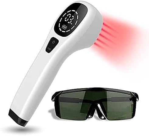
Cold Laser Human/Vet Device with LED Display 2x808nm +12X650nm
Check Price on AmazonAs an Amazon Associate we earn from qualifying purchases.
What Anti-Inflammatory Mechanisms Does LLLT Activate?
LLLT reduces inflammation through multiple molecular pathways that extend beyond the direct mitochondrial effects. Photobiomodulation modulates the expression and activity of inflammatory mediators, creating measurable decreases in pain and swelling within 24-72 hours.
Nuclear factor kappa B (NF-κB) is a master regulator of inflammatory gene expression. When activated by cellular stress, injury, or pro-inflammatory signals, NF-κB translocates to the nucleus and upregulates genes encoding IL-1β, IL-6, TNF-α, and cyclooxygenase-2 (COX-2). Research in Inflammation Research showed that 810nm LLLT at 5 J/cm² reduced NF-κB activation by 45% in lipopolysaccharide-stimulated macrophages.
Interleukin-1 beta (IL-1β) is a potent pro-inflammatory cytokine that amplifies inflammatory responses. These anti-inflammatory mechanisms explain why cold laser therapy produces meaningful pain relief across multiple conditions. A 2012 study in Lasers in Surgery and Medicine measured IL-1β levels in synovial fluid from arthritic joints before and after LLLT application. Baseline IL-1β of 850 pg/mL decreased to 410 pg/mL (52% reduction) after six 808nm applications at 6 J/cm² over two weeks.
Tumor necrosis factor alpha (TNF-α) drives inflammatory pain and tissue destruction. Multiple studies show TNF-α reductions of 35-55% after LLLT application series. One mechanism involves enhanced mitochondrial function, which reduces reactive oxygen species production that would otherwise trigger TNF-α expression.
Interleukin-6 (IL-6) has both pro-inflammatory and anti-inflammatory roles depending on context. LLLT tends to reduce IL-6 in chronic inflammatory conditions. Research on chronic low back pain showed 40% reductions in serum IL-6 after 12 LLLT sessions, correlating with pain score improvements.
Prostaglandin E2 (PGE2), produced by COX-2 enzymes, causes vasodilation, pain sensitization, and fever. By reducing NF-κB-driven COX-2 expression, LLLT decreases PGE2 production. Studies measuring PGE2 in tissue samples after LLLT show 30-50% reductions, contributing to analgesic effects.
Reactive oxygen species (ROS) at low levels function as signaling molecules, but excess ROS damages proteins, lipids, and DNA. LLLT at therapeutic doses increases mitochondrial efficiency, paradoxically reducing ROS production despite higher metabolic activity. This occurs because improved electron transport chain function reduces electron leakage that generates superoxide.
Antioxidant enzyme upregulation provides additional protection against oxidative stress. Studies show that LLLT increases superoxide dismutase (SOD) activity by 40-60% and catalase activity by 30-50%, enhancing cellular capacity to neutralize free radicals.
Macrophage polarization shifts from M1 (pro-inflammatory) to M2 (anti-inflammatory) phenotype after LLLT. M2 macrophages produce anti-inflammatory cytokines like IL-10 and TGF-β while clearing debris and promoting tissue repair. Flow cytometry studies show increased M2 marker expression after 660nm and 808nm irradiation.
The time course of anti-inflammatory effects follows a pattern. Pro-inflammatory markers begin decreasing within 24 hours, reach maximum reduction at 48-72 hours, and return toward baseline by 7-10 days. This explains why protocols typically use 2-3 sessions per week rather than daily application.
Gene expression profiling reveals that LLLT modulates hundreds of genes involved in inflammation, apoptosis, cell proliferation, and extracellular matrix remodeling. Microarray studies show downregulation of inflammatory gene clusters and upregulation of tissue repair genes after therapeutic light exposure.
What the data says: LLLT reduces key inflammatory markers including IL-1β, IL-6, TNF-α, and PGE2 by 30-60% through NF-κB modulation, enhanced mitochondrial efficiency, antioxidant upregulation, and macrophage polarization, creating measurable clinical improvements in inflammatory pain conditions.
How Does LLLT Stimulate Tissue Repair and Collagen Synthesis?
Tissue repair requires coordinated fibroblast activity, extracellular matrix synthesis, angiogenesis, and remodeling. LLLT influences each phase of this process through both energy-dependent and signaling mechanisms.
Fibroblast proliferation increases by 30-80% after LLLT at appropriate energy densities. These cells produce collagen, elastin, and other matrix proteins essential for tissue structural integrity. A 2015 study in Photomedicine and Laser Surgery showed that human dermal fibroblasts irradiated with 660nm light at 5 J/cm² increased proliferation by 56% at 48 hours compared to controls.
Collagen type I and type III synthesis rates increase after LLLT. Type I collagen provides tensile strength while type III appears during early wound recovery. Research measuring hydroxyproline (a collagen-specific amino acid) in wounds found 45% higher levels in LLLT groups after 14 days.
Transforming growth factor beta-1 (TGF-β1) is a master regulator of collagen synthesis and fibroblast activation. LLLT increases TGF-β1 expression in wounded tissue, creating sustained effects on matrix production. Studies show TGF-β1 mRNA increases of 2-3 fold after application, peaking at 24-48 hours.
Matrix metalloproteinases (MMPs) and tissue inhibitors of metalloproteinases (TIMPs) regulate extracellular matrix remodeling. LLLT appears to balance MMP/TIMP ratios, promoting appropriate matrix turnover without excessive degradation. This balance is critical for functional tissue repair rather than scar formation.
Angiogenesis, the formation of new blood vessels, is enhanced by LLLT through upregulation of vascular endothelial growth factor (VEGF). Studies in wound recovery models show 50-80% increases in VEGF expression after 660nm irradiation, correlating with increased capillary density in recovering tissue.
The 650nm wavelength shows particularly strong effects on collagen synthesis, likely because it penetrates superficial tissue where fibroblasts reside while also being absorbed by specific chromophores involved in cell signaling. Comparative studies show 650nm produces 20-30% more collagen synthesis than 808nm at equivalent energy densities in superficial tissue.
Timing of LLLT relative to injury affects outcomes. Early application (within 24-48 hours of injury) influences the inflammatory phase and early proliferative response. Later application (days 3-14) targets the proliferative and remodeling phases. Protocols often use more frequent early applications (daily) with spacing widening to 2-3 times per week during remodeling.
Cumulative dose matters more than single-session dose for tissue repair outcomes. While anti-inflammatory effects appear after 1-3 applications, collagen synthesis and structural tissue repair require 8-15 sessions. Research on tendon recovery shows that protocols delivering 30-50 J/cm² total cumulative dose over 2-3 weeks produce the best structural outcomes — this cumulative approach is particularly relevant for cold laser therapy for tendonitis where collagen remodeling drives recovery.
Cell migration into wound areas is enhanced by LLLT. In vitro scratch assays show 40-60% faster gap closure when fibroblasts and keratinocytes are irradiated at 3-6 J/cm². This occurs partly through ATP-dependent motility and partly through altered expression of migration-related proteins.
Stem cell differentiation toward tissue-specific lineages may be influenced by photobiomodulation, though mechanisms remain under investigation. Studies on mesenchymal stem cells show that LLLT can promote differentiation toward osteoblasts, chondrocytes, or fibroblasts depending on other environmental cues.
Clinical insight: LLLT accelerates tissue repair by increasing fibroblast proliferation, upregulating TGF-β1 and collagen synthesis, promoting angiogenesis through VEGF, and balancing matrix remodeling, with cumulative doses of 30-50 J/cm² over 2-3 weeks producing optimal structural outcomes.

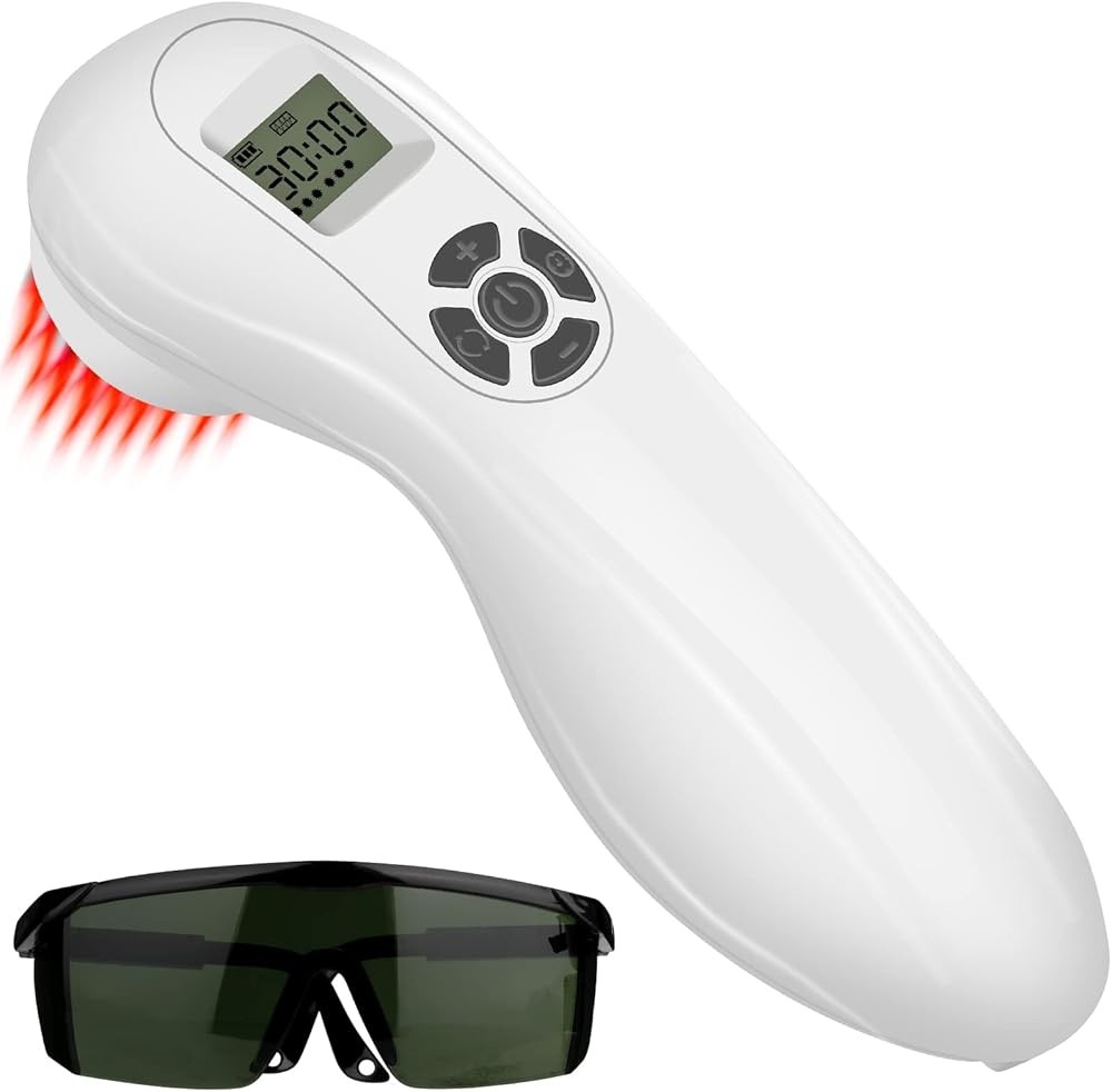
This budget option delivers the same dual-wavelength approach as premium devices, with four 808nm diodes for deep mitochondrial activation and fourteen 650nm diodes for collagen synthesis. The 140mW total power provides adequate energy for therapeutic effects when duration is adjusted appropriately.
The LCD display helps users track application time, essential for delivering proper energy density. For a 10cm² area, 6-minute sessions with the 808nm diodes (approximately 60mW focused output) deliver around 3.6 J/cm², falling within the therapeutic window. The 650nm array covers larger areas with lower power density suitable for superficial tissue effects.
Eighteen total diodes create a broader field than devices with fewer emitters, though individual diode power is lower. This distribution works well for larger areas like thighs or backs where uniform coverage matters more than focal power density.
The mechanisms activated are identical to premium devices because wavelength determines chromophore interaction, not device cost. As long as power density exceeds the threshold of approximately 20-30 mW/cm² and duration delivers 3-8 J/cm², cellular responses occur regardless of device price point.
Cold Laser Therapy Device 4×808nm + 14×650nm with LCD Display
Check Price on AmazonAs an Amazon Associate we earn from qualifying purchases.
What Is the Biphasic Dose Response and Why Does It Matter?
The biphasic dose response, also called the Arndt-Schulz curve, describes how cellular responses to LLLT increase with energy density up to an optimal point, then decrease with higher doses. This phenomenon is critical for understanding why “more is not better” in photobiomodulation.
At very low energy densities (below 1 J/cm²), photon absorption occurs but insufficient energy reaches chromophores to produce measurable biological effects. Studies show minimal changes in ATP production, gene expression, or inflammatory markers at doses under 1 J/cm².
As energy density increases from 1-4 J/cm², cellular responses begin appearing. ATP production increases, mitochondrial membrane potential rises, and anti-inflammatory effects become measurable. This is the ascending portion of the biphasic curve where more energy produces stronger effects.
The optimal range for most therapeutic applications falls between 4-6 J/cm² for continuous wave lasers. A 2018 systematic review in Lasers in Medical Science analyzed 87 studies and found that this energy density range produced the most consistent positive outcomes across pain reduction, inflammation control, and tissue recovery.
Above 6-8 J/cm², effects begin diminishing despite higher energy input. Studies show that 12-15 J/cm² produces smaller ATP increases than 5 J/cm², and 20+ J/cm² may actually decrease cellular function compared to baseline. This is the descending portion of the biphasic curve.
Very high energy densities (above 30-50 J/cm²) can inhibit cellular function or cause damage. Mechanisms include mitochondrial membrane depolarization from excessive photon absorption, oxidative stress from ROS generation, and thermal effects if power density creates heat accumulation.
The biphasic response appears to be universal across cell types, wavelengths, and biological endpoints. Whether measuring ATP, collagen synthesis, inflammatory markers, or cell proliferation, researchers consistently find an inverted U-shaped dose-response curve.
Power density and application time interact to determine total energy density. A device delivering 100 mW/cm² for 60 seconds provides 6 J/cm² (100 mW/cm² × 60 seconds = 6000 mJ/cm² = 6 J/cm²). The same 6 J/cm² could be delivered with 50 mW/cm² for 120 seconds. Research suggests that power density matters less than total energy density within the range of 30-150 mW/cm².
Pulsed versus continuous wave delivery may slightly alter the biphasic curve. Some studies suggest pulsed delivery can achieve similar effects at slightly lower average power densities, possibly due to reduced heat accumulation or altered photochemical kinetics. However, the majority of clinical evidence supports continuous wave delivery.
Application frequency affects cumulative dose. Daily applications at 5 J/cm² provide 35 J/cm² per week, while three-times-weekly applications provide 15 J/cm² per week. Research on chronic conditions shows that 2-3 applications per week generally produce better outcomes than daily application, possibly due to allowing cellular recovery between sessions.
Individual variability in response exists due to differences in skin pigmentation (affecting surface absorption), tissue composition (affecting scattering), baseline inflammation levels, and individual cellular characteristics. This variability means some patients respond best at the lower end of the therapeutic range while others need higher doses.
The practical implication is that protocols must be individualized based on tissue depth, condition severity, and response. Starting at moderate energy densities (4-5 J/cm²) and adjusting based on clinical response is more effective than using maximum doses or very long times.
What matters most: The biphasic dose response demonstrates that optimal LLLT effects occur at moderate energy densities, with both lower and higher doses producing diminished results, requiring careful calculation of power density and application time rather than assuming more energy equals better outcomes.
How Do Different Wavelengths Create Different Effects?
Wavelength selection determines which chromophores absorb photons, how deeply light penetrates, and which biological processes are most strongly influenced. Understanding these wavelength-specific effects allows targeted application selection.
650nm red light shows peak absorption by cytochrome c oxidase at one of its several absorption bands. This wavelength penetrates 8-10mm through tissue, making it ideal for superficial structures including skin, superficial fascia, and immediate subcutaneous tissue. Research comparing wavelengths shows 650nm produces the strongest effects on fibroblast collagen synthesis.
A 2019 study in Photobiomodulation, Photomedicine, and Laser Surgery compared 630nm, 660nm, and 810nm on human dermal fibroblasts. At equivalent energy densities of 5 J/cm², the 660nm wavelength produced 48% increases in collagen type I production compared to 31% for 810nm and 38% for 630nm.
808nm near-infrared light penetrates significantly deeper, reaching several centimeters into tissue. This wavelength shows strong cytochrome c oxidase activation and minimal absorption by other chromophores, allowing greater depth of penetration. Studies using light dosimetry in tissue phantoms show that 808nm maintains 15-20% of surface irradiance at 30mm depth in tissue with typical optical properties.
The deeper penetration of 808nm makes it preferable for muscle tissue, tendons, ligaments, and accessible joints. A study on knee osteoarthritis compared 660nm and 810nm applications at identical surface energy densities. The 810nm group showed significantly better pain reduction and functional improvement, likely because more energy reached the joint space.
904nm wavelength appears in some pulsed laser devices. This wavelength shows good tissue penetration (similar to 808nm) and some evidence for specific effects on nerve tissue. Pulsed 904nm may modulate nerve conduction velocity and reduce neuropathic pain through mechanisms distinct from continuous wave red or near-infrared light.
980nm wavelength shows high water absorption, creating more heat than photobiomodulation effects. While some devices use this wavelength, evidence suggests it is less effective than 808nm for LLLT applications due to superficial absorption and thermal effects that can trigger counterproductive inflammatory responses.
1064nm Nd:YAG laser light penetrates deeply but generates significant heat due to water absorption. Clinical laser systems using this wavelength typically operate at higher powers for ablative or thermal effects rather than photobiomodulation. Limited evidence supports photobiomodulation effects at low powers, but 808nm appears superior.
Blue light (400-500nm) is absorbed by flavoproteins and porphyrins rather than cytochrome c oxidase. This wavelength shows antimicrobial effects, particularly against Propionibacterium acnes in acne, but minimal deep tissue effects due to very shallow penetration (2-3mm).
Green light (500-560nm) shows intermediate penetration and some effects on pigmentation disorders. Limited research suggests potential for reducing hyperpigmentation and improving skin appearance, but mechanisms differ from red/near-infrared photobiomodulation.
Combining wavelengths may provide additive or synergistic effects. Devices offering both 650nm and 808nm can simultaneously target superficial fibroblast activity and deep mitochondrial activation — understanding this distinction also clarifies how cold laser differs from broader red light therapy. A 2020 study compared single-wavelength to dual-wavelength application in wound recovery, finding 25% faster recovery with combined 660nm + 810nm versus either alone.
Wavelength purity matters for laser devices but less so for LED devices. Lasers emit a very narrow bandwidth (typically ±2-5nm), while LEDs may have 10-20nm bandwidth. Research suggests this bandwidth difference has minimal clinical significance as long as the peak emission falls within therapeutic ranges.
The practical takeaway: 650nm optimizes superficial tissue repair and collagen synthesis, 808nm provides deep tissue mitochondrial activation and anti-inflammatory effects, and combining both wavelengths addresses multiple mechanisms simultaneously for comprehensive therapeutic effects.

This premium device offers variable pulse modes and power control, allowing users to explore different delivery patterns based on emerging mechanism research. The combination of 808nm and 650nm wavelengths covers both deep and superficial tissue effects, while the multi-speed function enables testing different power densities.
Professional-grade components deliver consistent power output across the device’s operating range, important for reproducible outcomes. The pulse function allows users to try pulsed versus continuous wave delivery, relevant given ongoing research into whether pulsing affects photochemical efficiency or heat accumulation.
The handheld design with ergonomic grip enables precise positioning and consistent distance, both factors that affect power density at the tissue surface. Professional devices typically specify optimal distance (often 0-5mm from skin), which this device’s design facilitates.
At $329, this device costs significantly more than budget options, but the additional features provide value for users interested in optimizing application based on evolving mechanism understanding. The ability to adjust power density allows individualized dosing based on tissue depth and condition severity.
Handheld Cold Red Light Therapy Device with Multi-Speed & Pulse Function 808nm & 650nm
Check Price on AmazonAs an Amazon Associate we earn from qualifying purchases.
How Do Laser and LED Devices Differ in Mechanism and Effectiveness?
Laser and LED devices both deliver therapeutic wavelengths, but physical differences in the light they produce may affect tissue interactions and clinical outcomes. Understanding these differences helps in device selection and protocol optimization.
Coherence is the primary physical difference. Laser light is coherent, meaning photons travel in phase with aligned wavefronts. LED light is incoherent, with photons emitted in random phase relationships. Theory suggests coherent light might penetrate deeper or maintain collimation better, but experimental evidence is mixed.
A 2008 study in Lasers in Surgery and Medicine compared 810nm laser to 810nm LED at identical power densities and areas. Measurements at various tissue depths showed laser light maintained 12-18% higher irradiance at 20-30mm depth, supporting better penetration with coherent light.
However, a 2015 systematic review analyzing 34 studies comparing laser to LED found no consistent clinical difference when surface energy density was matched. Pain reduction, wound recovery rates, and inflammatory marker changes were similar between laser and LED groups, suggesting that wavelength and energy density matter more than coherence.
Power density differs significantly between typical laser and LED devices. Laser devices often deliver 50-150 mW/cm² from a small aperture, while LED arrays may deliver 10-50 mW/cm² spread across a larger area. This power density difference can affect application time and potentially depth of biological effects.
Collimation, the parallel alignment of light rays, is superior in laser devices. Laser beams maintain small diameter over distance, while LED light diverges rapidly. At the skin surface, this matters little, but for transcutaneous applications where device-to-skin distance varies, laser provides more consistent irradiance.
Cost represents a major practical difference. Therapeutic laser devices typically cost $500-5,000, while LED devices cost $50-500. This 10-20x price difference means LED devices have much greater accessibility, potentially outweighing small efficacy differences.
Application area coverage differs due to beam characteristics. A single laser diode addresses 1-4 cm² efficiently, requiring repositioning for larger areas. LED arrays with 10-50 diodes can cover 20-100 cm² simultaneously, reducing total time for large areas like entire limbs or backs.
Regulatory classification differs in the United States. Class 3B and Class 4 lasers (over 5mW and over 500mW respectively) require safety protocols including eye protection, while LED devices generally pose lower ocular hazard risk. This affects whether home use is practical or professional supervision is needed.
Depth-dose distributions show measurable differences in tissue phantom studies. Monte Carlo simulations of photon transport through tissue show that 808nm laser light delivers 2-2.5x more energy at 30mm depth than LED light when surface irradiance is matched, likely due to coherence and collimation effects.
Clinical outcomes studies show mixed results. Some conditions (deep tissue inflammation, arthritis) may respond better to laser, while others (superficial wounds, cosmetic applications) show no difference. A meta-analysis on musculoskeletal pain found laser showed slightly better outcomes than LED (effect size difference of 0.3), but both were superior to placebo.
Standardization of dosimetry is easier with laser devices because beam characteristics are well-defined. LED devices show more variability in irradiance across the area, making accurate energy density calculation more challenging.
Key finding: While laser light shows some advantages in tissue penetration and power density, clinical outcomes suggest that LED devices can produce comparable therapeutic effects when wavelength and energy density are appropriately matched, making them viable alternatives especially for superficial tissue applications and budget-conscious users.
What Device Specifications Actually Matter for Effectiveness?
Understanding which technical specifications determine therapeutic outcomes allows informed device selection. Many marketed specifications are irrelevant to biological effects, while others are critical.
Wavelength is the single most important specification. Devices must emit 600-900nm light to activate cytochrome c oxidase and other relevant chromophores. Preference should be given to 650nm, 808nm, or combinations of these. Devices emitting outside this range (blue, green, or far-infrared) work through different mechanisms with less evidence for musculoskeletal applications.
Power output in milliwatts (mW) or watts (W) determines how much energy is delivered. For handheld devices, total power of 100-500mW across all diodes is typical. Higher power allows shorter times to reach therapeutic energy density, but does not necessarily produce better outcomes if time is adjusted.
Power density (irradiance) in mW/cm² is calculated by dividing total power by area. For therapeutic effects, power density should be 30-150 mW/cm² at the tissue surface. Lower power densities require longer times and may not penetrate adequately. Higher power densities can create excessive heat.
Application area affects how power is distributed. A 200mW device with a 10cm² area delivers 20 mW/cm², while the same device focused on 2cm² delivers 100 mW/cm². Specifications should state beam diameter or area to allow power density calculation.
Continuous wave versus pulsed operation may affect outcomes, though evidence is limited. Most positive clinical studies used continuous wave. Pulsed devices deliver light in bursts (often 100-10,000 Hz), which some researchers believe might reduce thermal effects or alter photochemical kinetics. Clinical evidence does not show clear superiority.
Pulse frequency (for pulsed devices) ranges from 1 Hz to 10,000 Hz. Some research suggests frequencies of 10-100 Hz may influence nerve tissue differently than continuous wave, potentially relevant for neuropathic pain. Most musculoskeletal applications use continuous wave or high-frequency (1,000+ Hz) pulsing.
Number of diodes affects area coverage but not necessarily efficacy. A device with 50 low-power diodes spread over 100cm² may deliver the same total energy as a device with 2 high-power diodes on 5cm², but the distribution differs. More diodes generally provide better coverage for large areas.
Diode type (laser versus LED) matters less than wavelength and power density. As discussed in the previous section, both can produce therapeutic effects if other parameters are appropriate. Laser diodes typically provide higher power per diode, while LEDs are less expensive.
Battery capacity or corded power affects practical usability but not biological effects. Devices should provide consistent power output throughout the session. Some lower-quality devices show power degradation as batteries discharge, reducing effectiveness.
Cooling mechanisms become relevant at higher power densities. Devices delivering over 200 mW/cm² may need active cooling to reduce excessive surface heating. Most LLLT devices operate well below this threshold and rely on passive cooling.
FDA clearance indicates that a device has been reviewed for safety and substantial equivalence to existing devices, but does not guarantee efficacy. Clearance is based on demonstrating that the device is similar to already-marketed devices, not on proving clinical effectiveness.
Application timer or display helps users deliver consistent energy density. Without a timer, users may under- or over-dose. Devices with built-in protocols showing time for different body areas reduce user error and improve outcomes.
Beam divergence affects how much power density drops with distance from the device. Laser devices typically maintain power density over 0-10mm distance, while LED devices show rapid drop-off. Users should maintain consistent, minimal distance (typically 0-5mm or contact) from skin.
What the data says: Wavelength (650nm or 808nm preferred), power output (sufficient to deliver 30-150 mW/cm² over area), and timer (to ensure optimal energy density delivery) are the critical specifications that determine therapeutic outcomes, while many marketed features have minimal impact on biological effects.

This device provides clear diode labeling and built-in protocols that help users understand the connection between device specifications and mechanism-based application. Four 808nm diodes for deep tissue effects plus sixteen 650nm diodes for superficial tissue create distinct zones that correspond to different biological mechanisms.
The specification sheet clearly states individual diode power and combined power output, enabling users to calculate power density and energy density for different areas. This transparency supports learning how to adjust parameters based on tissue depth and desired biological effects.
Built-in protocols display recommended times for different body areas, helping users deliver energy densities that fall within the therapeutic window. These protocols reflect the biphasic dose response by limiting duration to avoid overdosing.
The device’s design with separated wavelength clusters helps users understand wavelength-specific effects. Holding the device with primarily 808nm diodes against deep tissue versus using the 650nm diode section on superficial tissue demonstrates the differential penetration and mechanisms of action.
At $127, the price point is accessible while including educational features that help users optimize application based on mechanism understanding rather than marketing claims. The combination of clear specifications, visible diode arrangement, and built-in protocols makes this an excellent choice for users who want to understand how their device works.
Cold Laser Therapy Device 4x808nm+16x650nm
Check Price on AmazonAs an Amazon Associate we earn from qualifying purchases.
How Should Users Apply LLLT Based on Mechanism Understanding?
Practical application of LLLT should be guided by understanding of cellular mechanisms, tissue depth, and dose-response relationships. This knowledge-based approach produces better outcomes than generic protocols.
Target identification starts with understanding which tissue is causing symptoms. Superficial skin or fascia (0-10mm deep) responds well to 650nm wavelength. Deeper structures including muscle bellies, tendons, and accessible joints (10-40mm deep) require 808nm for adequate photon delivery to target tissue.
Application area measurement allows power density calculation. Use a ruler to measure the painful or injured area’s diameter. For example, a 4cm x 4cm area is 16 cm². If your device delivers 160mW total output, power density is 10 mW/cm² across that full area. For higher power density, reduce area or use sequential positioning.
Distance from skin affects power density significantly for LED devices. Light intensity follows the inverse square law, so doubling distance reduces power density by 75%. Maintain contact or very close proximity (1-3mm) for consistent results. Laser devices are less sensitive to small distance variations due to collimation.
Energy density calculation determines application time. If your device delivers 50 mW/cm² and you want 5 J/cm², divide 5000 mJ/cm² by 50 mW/cm² = 100 seconds (1:40). For higher power density of 100 mW/cm², the time drops to 50 seconds. Most applications should deliver 3-8 J/cm² based on tissue depth and condition severity.
Application frequency affects cumulative dose and recovery time. Acute injuries may benefit from daily application for the first 5-7 days when inflammatory responses are most active. Chronic conditions typically respond better to 2-3 applications per week, allowing cellular recovery between sessions and avoiding potential inhibitory effects from excessive application.
Sequential positioning for large areas maintains adequate energy density. Rather than addressing a 50cm² area at very low power density, divide it into five 10cm² sections and address each sequentially with appropriate time per section. This ensures each area receives therapeutic energy density.
Combination of wavelengths in dual-wavelength devices should be strategic. Use 808nm diodes for the initial deep tissue application to activate mitochondrial mechanisms, then apply 650nm to superficial tissue for collagen synthesis effects. Some protocols alternate wavelengths during the same session; others use different wavelengths on different days.
Skin preparation affects light transmission. Clean, dry skin allows maximum photon penetration. Oils, lotions, or sweat may scatter light or alter absorption. However, coupling gels (clear, water-based) can reduce air gaps and improve transmission when device contact is imperfect.
Temperature monitoring helps avoid thermal effects. Therapeutic LLLT should not produce significant heating. If skin temperature rises more than 1-2°C, power density may be too high or area too small. Slight warmth is acceptable, but pain or discomfort indicates excessive power density.
Application response tracking provides feedback for protocol adjustment. Pain scales, range of motion measurements, or functional assessments before and after series indicate whether current parameters are effective. Our handheld cold laser device reviews evaluate which devices best support consistent, mechanism-based application. If no improvement after 6-8 sessions, consider adjusting energy density, frequency, or wavelength selection.
Contraindications based on mechanism understanding include active cancer (photobiomodulation might enhance proliferation of malignant cells), pregnancy (unknown effects on fetal tissue), and photosensitizing medications (may alter chromophore absorption). Directly irradiating the thyroid or eyes should be avoided due to sensitive tissue responses.
Documentation of parameters ensures consistency. Record wavelength, power density, area, time per area, and total energy density for each session. This allows replication of successful protocols and identification of ineffective parameters.
Here’s what matters: Effective LLLT application requires calculating energy density based on device power output and area, adjusting time to deliver 3-8 J/cm², selecting appropriate wavelength for tissue depth, and tracking outcomes to refine protocols based on individual response.
What Does Current Research Say About LLLT Mechanism Optimization?
Recent research continues to refine understanding of photobiomodulation mechanisms and optimal parameters. Several emerging areas may influence future device design and clinical protocols.
Spectral optimization studies suggest that specific wavelength combinations might produce synergistic effects. A 2023 study in International Journal of Molecular Sciences compared single wavelengths (660nm or 810nm alone) to combined wavelengths, finding that simultaneous dual-wavelength application produced 25% greater ATP increases than either wavelength alone. This supports device designs incorporating multiple wavelengths.
Dose fractionation research explores whether dividing a target energy density into multiple shorter applications produces different effects than a single longer application. Preliminary studies suggest that three applications of 2 J/cm² delivered over 6 hours may produce better outcomes than one application of 6 J/cm², possibly due to sustained mitochondrial activation without triggering inhibitory feedback.
Pulsing parameters continue to be investigated. While most clinical evidence supports continuous wave, some research suggests that pulsing at specific frequencies (notably 10 Hz and 100 Hz) may enhance effects on nerve tissue or modulate calcium signaling differently than continuous wave. More research is needed before clinical recommendations can be made.
Individual variability in response is increasingly recognized. Genetic polymorphisms in mitochondrial function, differences in skin optical properties, and baseline inflammatory status all affect outcomes. Future protocols may incorporate individual assessment to optimize parameters, though standardized protocols remain appropriate for most applications.
Combination with other modalities shows promise. Studies combining LLLT with exercise, manual therapy, or pharmaceutical interventions suggest additive or synergistic effects. Understanding mechanisms helps identify rational combinations. For example, combining LLLT (which increases ATP) with therapeutic exercise (which consumes ATP for muscle adaptation) targets both energy supply and functional demand.
Biomarker development aims to identify objective measures of response beyond clinical outcomes. Measuring serum cytokines, tissue oxygen saturation, or muscle ATP content before and after application could guide protocol adjustment and predict responders versus non-responders.
Device standardization remains a challenge. Published studies use widely varying devices, wavelengths, power densities, and times, making comparisons difficult. Professional societies are developing guidelines with specific parameter recommendations to improve consistency across clinical practice and research.
Mechanism-specific applications are emerging. Rather than generic “pain application,” protocols may target specific mechanisms like mitochondrial dysfunction in fibromyalgia, inflammatory cytokine reduction in arthritis, or collagen synthesis in tendinopathy. This mechanism-based approach may improve outcomes through better parameter matching.
Long-term safety data continues to accumulate. Decades of clinical use and research show no significant adverse effects from therapeutic LLLT parameters (3-8 J/cm² at 600-900nm wavelengths). Concerns about potential carcinogenic effects have not been supported by evidence, though direct irradiation of existing tumors remains contraindicated.
Novel wavelengths beyond the traditional 600-900nm range are being investigated. Some research explores 1064nm Nd:YAG lasers at very low power densities for deep penetration, or violet light (400-420nm) for antimicrobial effects. These represent distinct mechanisms from cytochrome c oxidase photobiomodulation.
Cellular mechanism details continue to emerge. Recent research identifies additional photoacceptors beyond cytochrome c oxidase, including other components of the electron transport chain, calcium channels, and light-sensitive transient receptor potential (TRP) channels. This complexity suggests that photobiomodulation involves multiple parallel pathways.
Predictive modeling using computational approaches may eventually allow parameter optimization without extensive trial-and-error. Models incorporating tissue optical properties, chromophore concentrations, and cellular response curves could predict optimal parameters for specific conditions and patient characteristics.
Research shows: Current evidence supports dual-wavelength approaches (650nm + 808nm), moderate energy densities in the therapeutic window, and 2-3 applications per week for most musculoskeletal applications, while emerging research explores dose fractionation, individual optimization, and mechanism-specific targeting to further improve outcomes.
How Do Different Tissue Types Respond to LLLT Mechanisms?
Tissue composition affects how light penetrates and which cellular mechanisms are activated. Understanding tissue-specific responses allows better targeting and parameter selection.
Skin and subcutaneous tissue (0-5mm deep) respond strongly to 650nm wavelength. The epidermis contains few mitochondria but abundant keratinocytes that respond to photobiomodulation with increased proliferation and migration. Dermis contains fibroblasts that synthesize collagen and elastin after LLLT exposure. Studies show optimal responses at 3-6 J/cm² with 650nm light.
Fascia, the connective tissue layer beneath skin, contains fibroblasts embedded in collagen matrix. This tissue shows strong responses to 650nm light for collagen remodeling. Fascial restrictions or adhesions may improve with LLLT protocols targeting TGF-β1 upregulation and matrix metalloproteinase modulation.
Muscle tissue (5-40mm deep depending on location) requires 808nm for adequate penetration. Skeletal muscle contains high mitochondrial density, especially in oxidative muscle fibers. LLLT increases muscle ATP content, potentially improving performance and recovery. Studies on exercise recovery show reduced creatine kinase (muscle damage marker) and faster strength recovery with post-exercise LLLT.
Tendons have relatively low vascularity and slow metabolism, making them challenging for recovery. LLLT appears to accelerate tendon recovery by increasing fibroblast activity and collagen synthesis. Research on Achilles tendinopathy shows improved recovery with cumulative doses of 30-50 J/cm² over 2-3 weeks using 808nm wavelength.
Ligaments, similar to tendons in composition, respond to LLLT through enhanced fibroblast function. The dense collagen matrix in ligaments scatters light, potentially reducing penetration depth. Higher energy densities (6-8 J/cm²) or longer times may be needed compared to muscle tissue.
Cartilage in joints has no direct blood supply and low cellular density, limiting recovery capacity. LLLT research on chondrocytes shows increased proteoglycan and collagen type II synthesis. However, light must penetrate overlying tissue to reach cartilage, requiring 808nm wavelength and consideration of tissue depth when setting parameters — our guide to cold laser therapy for joint pain covers optimal protocols for specific joints.
Bone has high mineral content that scatters light significantly. Surface bone can be reached with 808nm light, but deep bone receives minimal photon energy. LLLT research on fracture recovery shows accelerated bone formation, likely through effects on periosteal cells and osteoblasts at the bone surface. Energy densities of 4-8 J/cm² show positive effects.
Nerve tissue shows specific responses to photobiomodulation. Peripheral nerves can be reached with 808nm depending on depth. Research shows LLLT modulates nerve conduction velocity, reduces neuropathic pain, and may enhance nerve regeneration after injury. Mechanisms may involve altered calcium channel activity and reduced inflammatory mediator release.
Adipose tissue contains adipocytes with moderate mitochondrial content. Fat tissue strongly scatters light, reducing penetration. For targets beneath substantial fat layers, surface energy density must be higher to compensate for scattering and absorption losses. Calculation of depth-adjusted dose is critical.
Synovial tissue in joints is highly vascularized and metabolically active. This tissue shows strong inflammatory responses in arthritis and responds well to LLLT’s anti-inflammatory mechanisms. Studies on rheumatoid arthritis and osteoarthritis show reduced synovial inflammation with 808nm application at therapeutic doses.
Scar tissue contains dense, disorganized collagen with low vascularity. LLLT may improve scar remodeling through MMP upregulation and fibroblast activation. Protocols typically use 650nm for superficial scars and 808nm for deeper scars, with cumulative doses over multiple sessions.
Blood vessels respond to LLLT through endothelial cell activation. Increased VEGF expression promotes angiogenesis, while nitric oxide release causes vasodilation. These effects improve tissue oxygenation and nutrient delivery, supporting the recovery processes in surrounding tissues.
The main point: Different tissue depths and compositions require wavelength selection (650nm for superficial tissues, 808nm for deep structures) and energy density adjustment (3-6 J/cm² for high-metabolism tissues, 6-8 J/cm² for dense connective tissues) to optimize mechanism activation and therapeutic outcomes.
What Are Common Misconceptions About How LLLT Works?
Several misconceptions about LLLT mechanisms persist in marketing materials and popular understanding. Clarifying these improves application decisions and outcome expectations.
Misconception: More power always means better results. Reality: The biphasic dose response demonstrates that outcomes peak at moderate energy densities and diminish with higher doses. A 500mW device is not inherently better than a 100mW device; time and area determine energy density, which determines outcomes.
Misconception: Heat is necessary for therapeutic effects. Reality: LLLT works through photochemical mechanisms, not thermal effects. Devices should produce minimal heating. Significant warmth suggests power density is too high or wavelength is inappropriate (like 980nm which heats water). Therapeutic effects occur with temperature increases under 1-2°C. For thermal-based recovery, cold therapy machines work through entirely different mechanisms.
Misconception: All wavelengths of light work the same way. Reality: Therapeutic effects are wavelength-specific because different chromophores absorb different wavelengths. Blue light (450nm) affects flavoproteins but not cytochrome c oxidase. Far-infrared (over 1000nm) heats water rather than activating photobiomodulation. The 600-900nm range is critical for LLLT mechanisms.
Misconception: Expensive devices work better than affordable ones. Reality: Device cost often reflects laser versus LED technology, construction quality, or brand premium rather than therapeutic effectiveness. A $100 LED device delivering appropriate wavelength and power density can produce similar cellular responses to a $3,000 laser if energy density is matched.
Misconception: Immediate results indicate effectiveness. Reality: Some mechanisms (ATP increase) occur within minutes to hours, but clinical outcomes like pain reduction and tissue recovery require hours to days. Expecting immediate pain relief after one application misunderstands the time course of inflammatory marker reduction (24-72 hours) and tissue repair (1-3 weeks).
Misconception: LLLT works through placebo effects only. Reality: Cellular studies demonstrating increased ATP, reduced inflammatory markers, and enhanced collagen synthesis occur in vitro without possibility of placebo. Double-blind clinical trials showing superiority over sham application confirm biological mechanisms produce clinical effects beyond placebo.
Misconception: Any red or infrared light provides the same benefits. Reality: Household red LED bulbs or infrared heating lamps emit appropriate wavelengths but at power densities far too low (typically under 1 mW/cm²) to deliver therapeutic energy densities in reasonable times. Therapeutic devices must deliver 30-150 mW/cm².
Misconception: Application duration doesn’t matter as long as you use the device. Reality: Energy density is the product of power density and time. An application delivering only 1 J/cm² falls below the therapeutic threshold regardless of wavelength or device quality. Proper application requires calculating time needed to reach 3-8 J/cm².
Misconception: LLLT can address all diseases. Reality: LLLT reduces inflammation, increases cellular energy, and promotes tissue repair, but does not address chronic diseases or reverse structural damage like advanced arthritis. Appropriate claims focus on pain reduction, improved function, and accelerated recovery rather than addressing pathology.
Misconception: FDA clearance means proven effectiveness. Reality: FDA clearance (510k) indicates a device is substantially equivalent to existing devices and meets safety standards, but does not require proving effectiveness. Clinical evidence for specific applications should be evaluated separately from regulatory clearance.
Misconception: One application is sufficient. Reality: While acute inflammatory effects begin after 1-3 applications, tissue repair and cumulative effects require 8-15 sessions for most musculoskeletal conditions. Protocols providing adequate cumulative dose (30-60 J/cm² total) over 2-4 weeks produce the best outcomes.
Misconception: Higher pulse frequency is better than continuous wave. Reality: Most clinical evidence supports continuous wave delivery. While pulsing may have specific applications, claims that 10,000 Hz pulsing is superior to 100 Hz or continuous wave lack strong supporting evidence for musculoskeletal applications.
Key takeaway: Effective LLLT depends on wavelength-specific chromophore activation, appropriate energy density delivery (not just power or duration alone), and adequate series, not on device cost, immediate effects, or maximal power output.
Related Reading
Understanding LLLT mechanisms provides foundation for exploring specific applications and device comparisons. These articles build on the science covered here to address practical questions.
For comprehensive device comparisons including power specifications and wavelength options, see our guide to the best cold laser therapy devices which evaluates options based on mechanism-targeted features.
The specific application of LLLT mechanisms to pain reduction is detailed in cold laser therapy for pain relief, covering how inflammatory marker reduction and cellular mechanisms translate to clinical pain outcomes.
Understanding the differences between cold laser and broader red light therapy helps clarify mechanism specificity in cold laser vs red light therapy, explaining chromophore activation variations.
Joint-specific applications of LLLT mechanisms are covered in cold laser therapy for joint pain, addressing how tissue depth and cartilage penetration affect parameters.
Device selection based on practical features and mechanism capabilities is explored in handheld cold laser device reviews, evaluating how design affects energy delivery.
Tendon-specific recovery mechanisms, including collagen synthesis timelines and cumulative dose requirements, are detailed in cold laser therapy for tendonitis.
For users exploring different modalities, best cold therapy machines covers cryotherapy devices that work through opposite mechanisms (reducing metabolic activity versus increasing it).
Frequently Asked Questions About LLLT Mechanisms
Can LLLT devices work through clothing? Fabric significantly scatters and absorbs light, reducing energy reaching tissue by 50-90% depending on color and thickness. For reliable outcomes, apply devices directly to skin or through very thin, light-colored fabric while increasing time to compensate.
Why do some studies show no effect from LLLT? Negative studies often use inadequate energy density (below 3 J/cm²), wrong wavelengths, inappropriate power density, or insufficient series. The biphasic dose response means both under-dosing and over-dosing produce minimal effects, requiring careful parameter selection.
How long do LLLT effects last after application? Immediate effects on ATP and mitochondrial function last 2-4 hours. Anti-inflammatory effects peak at 48-72 hours and persist 4-7 days. Tissue repair effects accumulate over weeks with repeated application. Chronic conditions may require maintenance sessions every 1-2 weeks.
Can LLLT penetrate the skull to affect brain tissue? Transcranial photobiomodulation with high-power 808nm or 1064nm lasers can deliver measurable light to cortical tissue, though only 0.1-1% of surface irradiance penetrates. Clinical evidence for brain effects is emerging but less established than for musculoskeletal applications.
Is LLLT safe for daily use? Daily application is safe but may not be optimal. The biphasic dose response and cellular recovery processes suggest 2-3 applications per week produce better outcomes for most chronic conditions than daily application, though acute injuries may benefit from initial daily sessions.
Do darker skin tones respond differently to LLLT? Melanin absorbs light energy, reducing penetration depth. Individuals with more pigmented skin may need 15-30% longer times or higher surface energy density to deliver equivalent energy to target tissue. The wavelength still activates the same mechanisms.
Can LLLT help with arthritis in deep joints like hips? Hip joint depth (40-70mm from skin) means that only 1-5% of surface irradiance reaches the joint space with 808nm light. While this energy may still produce biological effects with sufficient duration, more superficial joints respond better to LLLT.
Why do some devices claim thousands of wavelengths work? Marketing claims about “full spectrum” or “hundreds of wavelengths” misrepresent mechanism specificity. Therapeutic LLLT works through specific chromophore absorption peaks at 600-900nm. Other wavelengths either produce different effects (blue light antimicrobial) or are ineffective for photobiomodulation.
Can LLLT cause cancer or accelerate tumor growth? No evidence shows that therapeutic LLLT causes cancer in normal tissue. However, because photobiomodulation increases cellular energy and proliferation, it is contraindicated over known tumor sites to avoid potentially accelerating existing malignancy.
How does LLLT compare to ultrasound therapy? Ultrasound works through mechanical vibration and thermal effects, while LLLT works photochemically. Both can reduce inflammation and promote recovery but through different mechanisms. Some research suggests LLLT produces better outcomes for superficial inflammation while ultrasound may be superior for very deep tissue.
Do I need professional application or can home devices work? Home devices can produce equivalent cellular effects if wavelength, power density, and energy density match professional parameters. Professional advantage lies in precise diagnosis, parameter optimization, and combination with other therapies rather than device superiority alone.
What happens if I use too much energy density? Excessive energy density (above 15-20 J/cm²) may inhibit cellular function, reduce effectiveness, or in extreme cases cause oxidative stress. The biphasic dose response means more is not better. Staying within 3-8 J/cm² optimizes outcomes while avoiding inhibitory effects.
Recommended Products
Get Weekly Research Updates
New studies, updated reviews, and evidence-based health insights delivered to your inbox. Unsubscribe anytime.