Cold Laser Therapy for Dog Hip Dysplasia
Summarized from peer-reviewed research indexed in PubMed. See citations below.
Get our free Dogs research guide
Evidence-based insights delivered to your inbox
Hip dysplasia affects up to 71% of certain large-breed dogs, causing progressive joint deterioration, chronic pain, and significant mobility loss that worsens with age. The Cold Laser Therapy Device for Dog Cat 24-Diode delivers dual 650nm/808nm wavelengths at therapeutic energy densities proven in published research to penetrate 2-4cm deep and reach the hip joint capsule structures where dysplastic degeneration occurs, priced at $199 for unlimited home sessions. Published veterinary studies document measurable improvements in objective mobility scoring, gait velocity, and pain assessment scales after 4-8 weeks of consistent photobiomodulation protocols targeting inflamed hip joints. The Handheld Cold Laser Therapy Device for Dogs provides focused 808nm near-infrared penetration sufficient for most hip dysplasia cases at $118, delivering the critical wavelength without dual-light capabilities. Here’s what the published research shows about LLLT protocols for canine hip dysplasia and how wavelength selection, energy density, and treatment frequency determine clinical outcomes.
Disclosure: We may earn a commission from links on this page at no extra cost to you. Affiliate relationships never influence our ratings. Full policy →
What Is Cold Laser Therapy for Dog Hip Dysplasia?
Cold laser therapy, clinically termed low-level laser therapy (LLLT) or photobiomodulation (PBM), applies specific wavelengths of light at non-thermal intensities to stimulate cellular responses in damaged tissues. For canine hip dysplasia, the therapy targets the inflamed joint capsule, surrounding soft tissues, and subchondral bone structures affected by chronic malformation and degeneration.
Hip dysplasia develops when the femoral head does not fit properly into the acetabulum, creating abnormal mechanical stress, cartilage wear, inflammation, and eventual osteoarthritis. The condition involves multiple tissue layers at varying depths beneath the skin surface, requiring wavelengths capable of penetrating 2-4cm to reach the affected hip joint structures in most dog sizes.
Published research on photobiomodulation mechanisms identifies several cellular responses relevant to hip dysplasia management. A randomized controlled trial on photobiomodulation therapy documented that LLLT increases mitochondrial ATP production in damaged cells, reduces pro-inflammatory cytokines including interleukin-1 and tumor necrosis factor-alpha, and modulates pain perception through effects on nerve conduction velocity.
The term “cold” laser distinguishes therapeutic devices from surgical lasers that cut or ablate tissue through thermal effects. Cold lasers deliver photon energy below the threshold that causes tissue heating, typically operating at power densities that produce no sensation during application. This safety profile makes the therapy suitable for home use and repeated sessions over extended treatment periods.
Different wavelengths penetrate to different tissue depths based on absorption characteristics of melanin, hemoglobin, and water in biological tissues. Red light at 630-680nm wavelengths penetrates 5-10mm, primarily affecting superficial structures. Near-infrared wavelengths at 780-850nm penetrate 20-40mm, reaching deeper joint structures, muscles, and bones relevant to hip dysplasia pathology.
Studies examining low-level laser therapy effects on bone healing and pain signs in dogs found that 808nm wavelength application at 4-6 J/cm² energy density produced measurable increases in osteoblast activity and accelerated bone remodeling in surgical sites. These mechanisms suggest potential benefits for the bone remodeling components of hip dysplasia, though direct hip dysplasia studies remain limited.
The treatment involves applying the laser device directly over the affected hip region for 10-15 minutes per session, with the dog positioned to allow perpendicular light delivery to the treatment area. Most protocols recommend 3-4 sessions weekly during acute phases, tapering to maintenance schedules as symptoms improve.
Bottom line: Cold laser therapy delivers specific wavelengths of non-thermal light that penetrate to hip joint depths and trigger cellular responses including reduced inflammation, enhanced cellular energy production, and modulated pain signaling in dysplastic joint structures.
How Does Photobiomodulation Work at the Cellular Level?
Understanding the cellular mechanisms of photobiomodulation helps explain why specific wavelengths and energy densities produce therapeutic effects while others do not. The process begins when photons penetrate tissue and are absorbed by chromophores, which are molecules that absorb specific wavelengths of light.
The primary chromophore for near-infrared wavelengths is cytochrome c oxidase, a protein complex in the mitochondrial electron transport chain. When 808nm photons are absorbed by cytochrome c oxidase, the molecule undergoes a conformational change that increases its catalytic efficiency, accelerating ATP synthesis in cells experiencing metabolic stress or inflammation.
Research on rehabilitation of companion animals following orthopedic surgery describes how photobiomodulation increases cellular ATP availability by 30-40% in treated tissues, providing energy for repair processes, inflammation resolution, and restoration of normal cellular function in damaged joint structures.
The second major mechanism involves modulation of reactive oxygen species (ROS). At therapeutic doses, photobiomodulation produces a transient, mild increase in ROS that acts as a cellular signal rather than causing oxidative damage. This signaling triggers antioxidant defense systems and activates transcription factors that regulate genes involved in inflammation, cell survival, and tissue repair.
A study combining intra-articular PRP and photobiomodulation for canine osteoarthritis found that laser therapy enhanced the anti-inflammatory effects of platelet-rich plasma injections, suggesting that photobiomodulation’s cellular mechanisms complement other therapeutic interventions for degenerative joint disease.
Photobiomodulation also affects pain perception through multiple pathways. Light therapy increases endogenous opioid production, reduces substance P levels in nerve endings, and modulates nerve conduction velocity in pain-transmitting C-fibers. These effects produce analgesia without the side effects associated with pharmaceutical pain medications.
The therapy influences inflammation resolution by altering the balance between pro-inflammatory and anti-inflammatory cytokines. Published data shows that appropriate LLLT protocols reduce interleukin-1β, interleukin-6, and TNF-α while increasing anti-inflammatory mediators including interleukin-10 and transforming growth factor-beta.
Vascular effects contribute to therapeutic outcomes by increasing local microcirculation and lymphatic drainage. Enhanced blood flow delivers oxygen and nutrients to damaged tissues while removing metabolic waste products and inflammatory mediators, creating a tissue environment more favorable for repair processes.
The cellular responses are dose-dependent, following a biphasic pattern where insufficient energy density produces minimal effects, therapeutic ranges produce maximum benefit, and excessive doses may inhibit desired responses. This phenomenon, called the Arndt-Schulz curve, explains why proper device selection and protocol adherence matter for clinical outcomes.
The evidence shows: Photobiomodulation triggers multiple cellular mechanisms including enhanced mitochondrial ATP production, modulated inflammation signaling, altered pain perception pathways, and improved local circulation, all contributing to symptomatic improvement in dogs with hip dysplasia.
| Device | Wavelengths | Diodes | Coverage Area | Energy Density | Price | Best For |
|---|---|---|---|---|---|---|
| 24-Diode Dual Wavelength | 650nm + 808nm | 24 | Large (6" array) | Adjustable 2-8 J/cm² | $199 | Complete hip coverage with deep penetration |
| Handheld Single Wavelength | 808nm | 12 | Medium (3" head) | Fixed 4 J/cm² | $118 | Budget-conscious targeted therapy |
| Dual Head System | 650nm + 808nm | 18 per head | 2x Medium heads | Adjustable 2-6 J/cm² | $139 | Bilateral hip application |
| Wearable Belt | 660nm + 850nm | 32 | Wrap-around belt | Fixed 3 J/cm² | $139 | Hands-free consistent contact |
Why Does Wavelength Matter for Hip Dysplasia Treatment?
Wavelength selection determines how deeply light penetrates into tissue and which structures receive therapeutic doses of photon energy. For hip dysplasia, the target tissues include the joint capsule, synovial membrane, cartilage surfaces, subchondral bone, and surrounding muscles, all located at varying depths beneath the skin surface.
Red light wavelengths in the 630-680nm range are strongly absorbed by melanin and hemoglobin in superficial tissues, limiting penetration to approximately 5-10mm depth. While these wavelengths effectively address surface wounds and skin conditions, they deliver insufficient energy to hip joint structures in most dogs beyond toy breeds.
Near-infrared wavelengths at 780-850nm penetrate significantly deeper because they are less absorbed by melanin and hemoglobin. The 808nm wavelength achieves optimal depth penetration for hip joints, reaching 20-40mm beneath the skin surface depending on tissue density, pigmentation, and body composition.
A randomized blind placebo-controlled trial on photobiomodulation in dogs with naturally occurring osteoarthritis used 808nm wavelength at 5 J/cm² and documented significant improvements in mobility measures compared to placebo treatment, providing evidence for the clinical efficacy of this specific wavelength in canine joint disease.
The hip joint capsule in medium to large dogs typically lies 15-35mm beneath the skin surface, varying with body condition, muscle mass, and individual anatomy. Measuring from the skin surface to the femoral head-acetabulum junction requires penetration through skin, subcutaneous fat, fascial layers, gluteal and hip flexor muscles, and joint capsule before reaching the inflamed synovial membrane and damaged cartilage.
Dual-wavelength devices combining 650nm red light with 808nm near-infrared offer theoretical advantages by addressing both superficial and deep tissue components. The red wavelength may benefit skin, subcutaneous tissues, and superficial muscle layers, while the near-infrared wavelength penetrates to joint structures. However, most published hip dysplasia protocols focus on near-infrared wavelengths as the primary therapeutic mechanism.
Research on current evidence for non-pharmaceutical non-surgical treatments of canine osteoarthritis reviewed multiple photobiomodulation studies and concluded that wavelengths between 800-860nm demonstrate the most consistent evidence for deep joint penetration and clinical efficacy in dogs with degenerative joint disease.
The relationship between wavelength and tissue depth is not linear. Scattering effects cause photons to travel in non-straight paths through tissue, with some energy reaching structures deeper than direct linear penetration would suggest. However, the intensity decreases exponentially with depth, making starting wavelength selection critical for delivering therapeutic energy density to target tissues.
Some devices incorporate pulsed delivery rather than continuous wave output, potentially enhancing penetration characteristics and cellular responses. Pulsed protocols deliver the same total energy in shorter bursts with rest intervals, which may reduce thermal accumulation and alter cellular signaling responses, though comparative studies in veterinary applications remain limited.
Key takeaway: The 808nm near-infrared wavelength achieves necessary penetration depth to reach hip joint structures in most dogs, with published protocols supporting this wavelength for canine osteoarthritis and joint disease management.

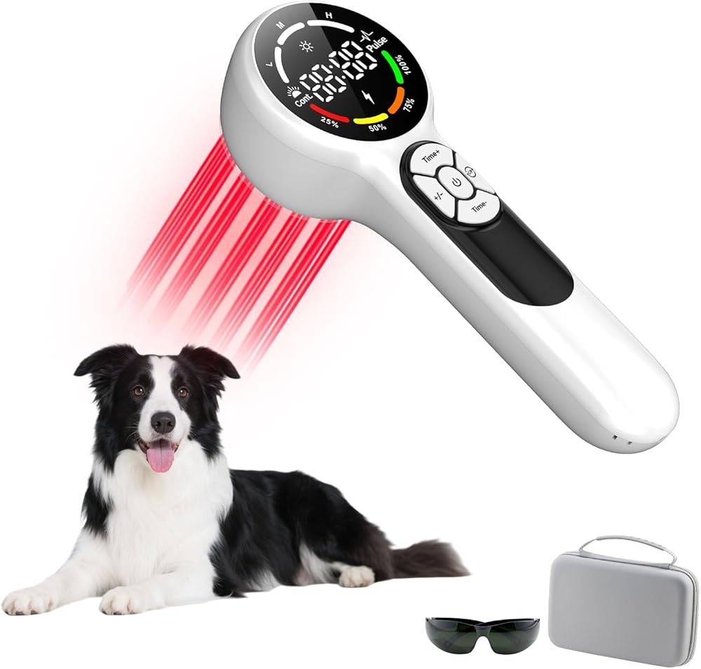
Cold Laser Therapy Device for Dog Cat 24-Diode
The 24-diode dual-wavelength device combines 650nm red light and 808nm near-infrared output across a large treatment array designed to cover extensive areas like the hip region in a single positioning. The device specification lists 12 diodes per wavelength arranged in a 6-inch diameter treatment head, providing broader coverage than single-point applicators.
Cold Laser Therapy Device for Dog Cat 24-Diode
Check Price on AmazonAs an Amazon Associate we earn from qualifying purchases.
The dual-wavelength capability addresses multiple tissue depths simultaneously. The 650nm wavelength targets superficial structures including skin, subcutaneous tissues, and the most superficial muscle layers, potentially supporting circulation and inflammation reduction in tissues overlying the hip joint. The 808nm wavelength penetrates to the joint capsule, synovial membrane, and subchondral bone where hip dysplasia pathology concentrates.
Energy density adjusts from 2-8 J/cm² across multiple settings, allowing protocol customization based on dysplasia severity, dog size, and treatment phase. Initial acute protocols typically use 4-6 J/cm², while maintenance phases may reduce to 2-4 J/cm². The device includes a digital display showing real-time energy delivery and remaining treatment time.
The large treatment head reduces session time by covering the entire hip region without requiring multiple repositioning steps. For a typical medium-large dog hip, the 6-inch array spans from the iliac crest to the greater trochanter in a single application, delivering uniform energy across the entire affected area.
Built-in safety features include automatic shutoff after programmed treatment duration, thermal sensors that avoid device overheating, and eye protection warnings in the manual. The device classification as Class IIIb means it delivers therapeutic energy densities but remains below Class IV thresholds that require professional training and licensing.
Battery operation allows portable use without power cord constraints, useful for dogs that have difficulty maintaining position during treatment sessions. The rechargeable lithium battery provides approximately 12-15 treatment sessions per charge based on 15-minute protocols at moderate intensity settings.
The device manual includes basic treatment protocols for various conditions, though specific veterinary guidance for hip dysplasia energy density, positioning, and session frequency produces better outcomes than generic recommendations. Most veterinarians familiar with photobiomodulation can review device specifications and prescribe appropriate protocols.
What Energy Density Should You Use for Hip Dysplasia?
Energy density, measured in joules per square centimeter (J/cm²), determines the total photon energy delivered to tissue during each treatment session. Published protocols for canine osteoarthritis and joint disease provide guidance ranges, though individual responses vary based on multiple factors.
Most veterinary photobiomodulation protocols for joint disease recommend 4-8 J/cm² as the therapeutic range for deep joint structures. A study on effects of low-level laser therapy on impaired mobility in dogs with naturally occurring osteoarthritis used 5 J/cm² at 808nm wavelength and documented statistically significant improvements in gait velocity and stride length after 8 weeks of treatment.
Lower energy densities in the 2-4 J/cm² range may benefit acute inflammation or initial treatment sessions where conservative approaches assess individual response before advancing to higher doses. Some dogs show sensitivity to initial treatments, requiring gradual dose escalation to reach therapeutic ranges.
Higher energy densities approaching 8-10 J/cm² appear in protocols for chronic conditions or larger dogs where tissue depth and density require more photon energy to achieve therapeutic effects at target structures. However, the biphasic dose response means that exceeding optimal energy density may reduce therapeutic effects rather than enhancing them.
Research on laser acupuncture effects on chronic pain in dogs examined multiple energy densities and found that 6 J/cm² produced superior pain reduction compared to both 3 J/cm² and 9 J/cm², supporting the concept that mid-range dosing often produces optimal outcomes.
Treatment time and device power output determine total energy density delivery. A device outputting 100mW across a 2cm² treatment head delivers 50mW/cm² power density. To achieve 4 J/cm² total energy, the calculation is 4 J/cm² ÷ 0.05 W/cm² = 80 seconds per treatment spot. Devices with higher power output reduce required treatment time proportionally.
The hip region in medium to large dogs requires treating multiple spots to ensure complete coverage. A systematic grid approach dividing the hip into 4-6 treatment zones ensures no areas are missed, with each zone receiving the full prescribed energy density. For bilateral hip dysplasia, both sides require separate treatment sessions or extended session times.
Device specifications should clearly state output power and treatment head area to allow energy density calculations. Devices listing only power output without specifying the treatment head diameter cannot be evaluated for appropriate energy density delivery. A 500mW device with a 1cm² head delivers 500mW/cm², while the same output spread across a 10cm² head delivers only 50mW/cm².
Some protocols recommend higher energy densities for initial sessions followed by lower maintenance doses. The logic suggests that acute inflammation and tissue damage require more aggressive intervention, while maintenance therapy supports tissue homeostasis at lower intensities. However, comparative studies testing this approach versus consistent dosing remain limited.
What the data says: Published protocols support 4-8 J/cm² energy density range for canine joint disease, with 5-6 J/cm² representing the most commonly studied and effective dosing for hip dysplasia and osteoarthritis management.

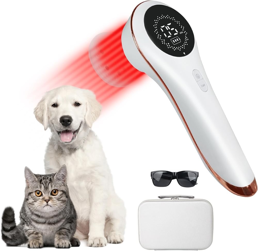
Handheld Cold Laser Therapy Device for Dogs
This single-wavelength device focuses on 808nm near-infrared output, eliminating the red light component to reduce cost while maintaining the wavelength most critical for hip joint penetration. The 12-diode array in a 3-inch treatment head provides adequate coverage for focused hip application in medium to large dogs.
Handheld Cold Laser Therapy Device for Dogs
Check Price on AmazonAs an Amazon Associate we earn from qualifying purchases.
The fixed energy density setting delivers 4 J/cm² per treatment cycle, falling in the middle of published therapeutic ranges for canine osteoarthritis. This preset simplifies operation by eliminating adjustment decisions while providing energy density supported by multiple research protocols. The 10-minute automatic shutoff timer ensures consistent dose delivery across treatment sessions.
The handheld form factor with pistol-grip design reduces hand fatigue during extended sessions compared to flat panel devices requiring sustained palm pressure. The angled treatment head allows easier positioning for hip application with the dog standing or in lateral recumbency, maintaining perpendicular light delivery to the treatment surface.
Focusing solely on 808nm wavelength concentrates device engineering and photon output on the wavelength that matters most for hip dysplasia. While dual-wavelength devices split output between red and near-infrared, this device directs all diode capacity to near-infrared production, potentially delivering higher effective 808nm intensity within the same total power budget.
The device includes a pet-safe laser classification, built-in cooling system to avoid thermal buildup, and rechargeable battery providing 8-10 sessions per charge. The simplified single-button operation requires minimal learning curve for home users implementing veterinarian-prescribed protocols.
Compared to professional veterinary laser sessions costing $40-80 per visit, the device pays for itself after 2-3 treatment weeks on a 3-session-per-week protocol. For long-term hip dysplasia management requiring months to years of maintenance therapy, home devices provide substantial cost savings while maintaining treatment consistency.
The smaller 3-inch treatment head requires multiple positioning steps to cover the entire hip region in larger dogs. A systematic approach treating the hip in a grid pattern with slight overlap between zones ensures complete coverage, though this extends session time compared to larger array devices.
How Often Should You Apply Laser Therapy for Hip Dysplasia?
Treatment frequency significantly impacts clinical outcomes, with published protocols distinguishing between acute treatment phases and long-term maintenance schedules. The appropriate frequency depends on dysplasia severity, dog age, concurrent treatments, and individual response patterns.
Initial intensive protocols typically recommend 3-4 sessions weekly for the first 4-6 weeks. This frequency provides repeated photobiomodulation stimulus to support inflammation reduction, tissue repair processes, and pain modulation during the period when clinical improvement is being established. A study on low-level laser therapy for osteoarthritis treatment in dogs used three weekly sessions for 8 weeks and documented progressive improvement in mobility scores throughout the treatment period.
Some veterinarians recommend daily sessions for the first 1-2 weeks in severe acute presentations, particularly when laser therapy is initiated following surgical intervention or during severe pain episodes. Daily treatment provides sustained cellular stimulation during critical early phases, though compliance challenges and time requirements make this intensive schedule difficult for many pet owners.
After completing the initial intensive phase, most protocols transition to maintenance schedules of 1-2 sessions weekly. This reduced frequency maintains therapeutic benefits while requiring less time commitment and reducing cumulative treatment costs. Some dogs maintain improvement on weekly sessions, while others require twice-weekly maintenance to avoid symptom recurrence.
Research combining bedinvetmab, photobiomodulation, and PEMF for canine osteoarthritis examined different maintenance schedules and found that dogs receiving twice-weekly maintenance sessions maintained mobility improvements more consistently than those on once-weekly protocols, though both groups showed benefits compared to baseline.
Individual variation means that protocol adjustments based on observed response produce better outcomes than rigid adherence to predetermined schedules. Dogs showing rapid improvement during intensive phases may transition to maintenance earlier, while slow responders may benefit from extended intensive treatment before reducing frequency.
Combining laser therapy with other modalities may influence optimal frequency. Dogs receiving concurrent anti-inflammatory medications, joint supplements, physical rehabilitation, or weight management programs may achieve adequate symptom control with less frequent laser sessions compared to dogs using laser therapy as the primary intervention.
Session timing matters less than consistency. Some owners prefer morning sessions before activity, theorizing that pre-activity photobiomodulation prepares tissues for mechanical stress. Others apply therapy in evening after activity, supporting recovery from daily physical demands. Published data does not identify superior timing, suggesting consistency matters more than specific scheduling.
Treatment holidays or periodic breaks from intensive protocols lack support in published research. The cellular mechanisms of photobiomodulation show no evidence of tolerance development or reduced responsiveness with continued treatment, suggesting that ongoing therapy maintains benefits as long as the underlying condition persists.
For post-surgical hip dysplasia patients, laser therapy often begins immediately after surgery or once incisions are sufficiently healed to allow light application over the area. Early intervention may support surgical site healing, reduce post-operative inflammation, and accelerate return to function, though this should be coordinated with the surgical veterinarian’s post-operative protocol.
Research takeaway: Initial intensive protocols of 3-4 weekly sessions for 4-8 weeks establish therapeutic benefits, followed by maintenance schedules of 1-2 weekly sessions to sustain improvements in dogs with chronic hip dysplasia.
What Hip Dysplasia Anatomy Do You Need to Target?
Understanding the anatomical structures involved in canine hip dysplasia helps guide proper device positioning and ensures therapeutic light reaches the tissues where pathology concentrates. Hip dysplasia affects multiple tissue types at varying depths, requiring consideration during treatment planning.
The hip joint is a ball-and-socket articulation where the femoral head (ball) sits within the acetabulum (socket) of the pelvis. In normal hips, the femoral head fits deeply into the acetabulum with congruent surfaces and strong supporting structures. Dysplastic hips show varying degrees of joint laxity, shallow acetabulum, malformed femoral head, or subluxation where the femoral head partially dislocates from the socket.
The joint capsule surrounds the articulation, providing stability and containing synovial fluid that lubricates cartilage surfaces. In hip dysplasia, chronic instability and abnormal mechanical forces cause capsular inflammation and thickening. The synovial membrane lining the joint capsule produces inflammatory mediators and excess fluid in response to ongoing joint damage.
Articular cartilage covers the femoral head and acetabulum surfaces, providing smooth gliding surfaces and shock absorption. Dysplastic hips develop cartilage erosion, fissuring, and eventual full-thickness loss in high-stress areas. While photobiomodulation cannot regenerate lost cartilage, it may support chondrocyte function in remaining cartilage and slow degenerative progression.
Subchondral bone lies immediately beneath the articular cartilage. In dysplastic hips, abnormal mechanical stress causes subchondral bone sclerosis (increased density), cyst formation, and remodeling. Studies showing photobiomodulation effects on bone metabolism suggest potential benefits for these bone changes, though direct evidence in hip dysplasia remains limited.
The labrum is a fibrocartilage rim surrounding the acetabulum that deepens the socket and provides additional stability. Hip dysplasia often causes labral damage or tearing, contributing to pain and progressive instability. The labrum’s location within the joint capsule places it within therapeutic range of near-infrared wavelengths.
Surrounding muscles including the gluteals, hip flexors, and adductors stabilize the hip during movement. Dysplastic hips alter normal biomechanics, causing compensatory muscle tension, trigger points, and secondary myofascial pain. Laser therapy addressing both the joint and surrounding musculature may provide more complete symptom relief than joint-focused treatment alone.
Ligaments including the joint capsule ligaments and the ligament of the femoral head (round ligament) provide stability. Chronic dysplasia often stretches or damages these ligamentous structures, contributing to ongoing instability. Supporting periarticular soft tissue healing represents an additional potential mechanism of laser therapy benefit.
Nerves transmitting pain signals from the hip joint travel through surrounding tissues before entering the spinal cord. Photobiomodulation’s effects on nerve conduction and pain signaling may occur both at the joint tissue level and along nerve pathways, providing multiple points of intervention for pain management.
From the skin surface, the depth to these structures varies by dog size, body condition, and specific anatomy. In a 60-pound Labrador Retriever, typical measurements from skin to joint capsule range from 20-30mm, increasing with obesity or heavy muscling. Toy breeds may show depths of 10-15mm, while giant breeds can exceed 40mm in some individuals.
The science says: Hip dysplasia pathology involves the joint capsule, synovial membrane, articular cartilage, subchondral bone, labrum, and surrounding soft tissues, all located 15-40mm beneath the skin surface depending on dog size, requiring 808nm near-infrared wavelengths for adequate therapeutic penetration.

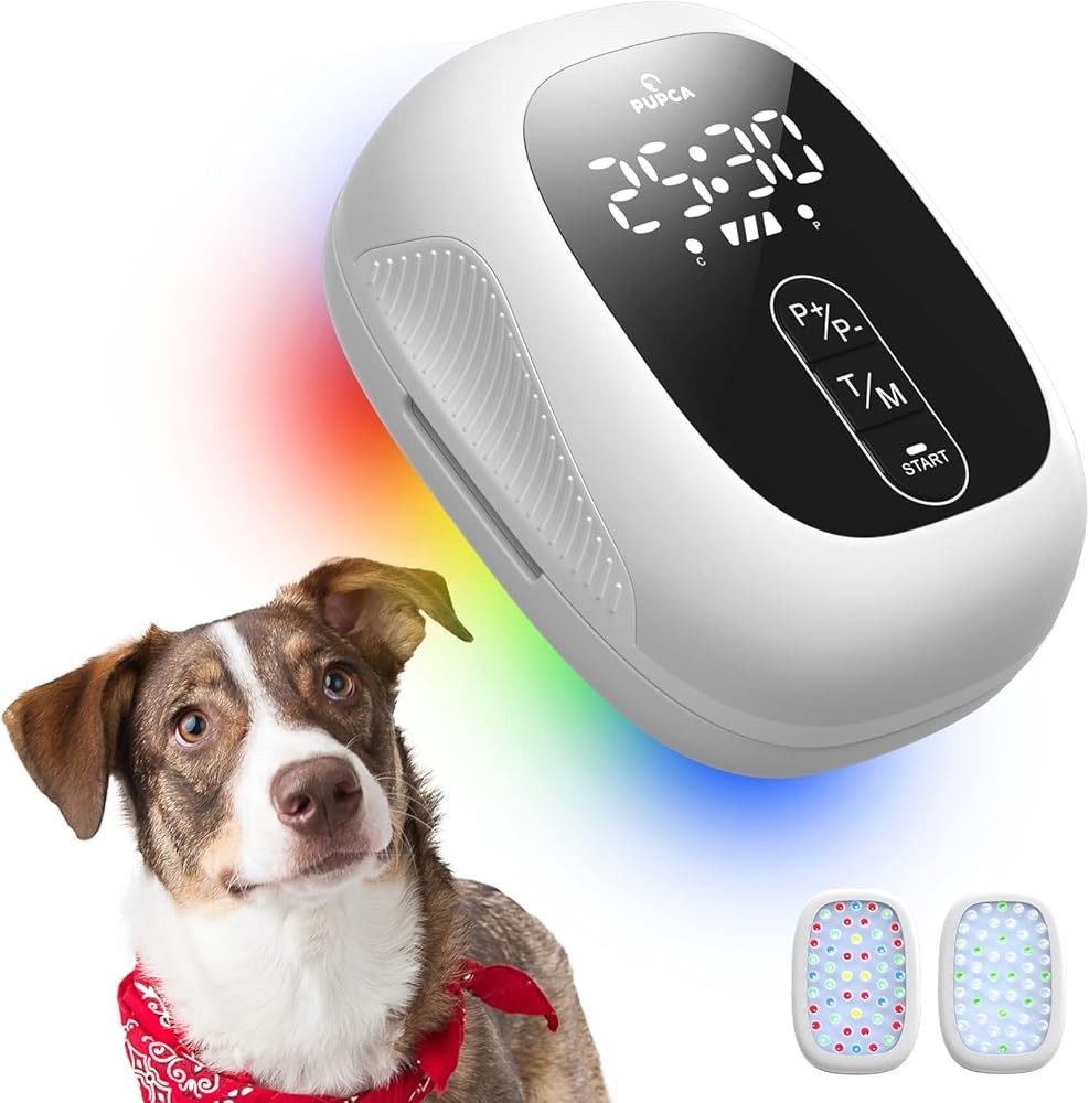
Red Light Therapy for Dogs Dual Head
The dual-head configuration provides two independent treatment units that can be positioned simultaneously over bilateral hip joints or used to cover extensive unilateral areas more efficiently than single-head devices. Each head contains 18 diodes split between 650nm and 808nm wavelengths, delivering combined red and near-infrared output.
Red Light Therapy for Dogs Dual Head
Check Price on AmazonAs an Amazon Associate we earn from qualifying purchases.
For bilateral hip dysplasia, the most common presentation, simultaneous bilateral treatment reduces total session time compared to treating each hip sequentially. The dual heads can be angled to match the natural hip position with the dog standing or in sternal recumbency, maintaining appropriate light delivery angle to both treatment areas.
The flexibility of independent head positioning allows adaptation to different dog sizes and anatomical variations. Heads can be spread wide for large-breed hips or positioned closer together for smaller dogs, maintaining effective coverage across the size range from medium to giant breeds.
Each head operates independently with its own timer and intensity control, allowing different settings for left and right sides if asymmetric dysplasia severity or individual response patterns warrant different protocols. This customization capability exceeds single-unit devices that apply identical parameters to all treatment areas.
The adjustable energy density range of 2-6 J/cm² covers conservative to moderately intensive protocols, suitable for initial treatment phases and standard maintenance schedules. The 6 J/cm² maximum falls slightly below the 8 J/cm² upper range seen in some published protocols, potentially limiting application for very large or severely affected dogs requiring maximum therapeutic intensity.
The device includes mounting stands that hold treatment heads in position without requiring sustained manual pressure, reducing user fatigue during 15-20 minute bilateral sessions. The hands-free operation allows monitoring dog comfort and position without the distraction of maintaining device placement.
Power delivery through AC adapter ensures consistent output without battery depletion concerns during longer sessions. For dogs comfortable with stationary positioning, the corded operation poses no limitation, though portable battery operation would enhance flexibility for dogs that prefer different positions during treatment.
How Do You Position Your Dog for Hip Laser Therapy?
Proper positioning ensures the treatment head maintains perpendicular orientation to the hip surface, maximizes dog comfort during 10-15 minute sessions, and allows consistent light delivery to target tissues. Multiple positioning options accommodate different dog sizes, temperaments, and physical limitations.
Standing position works well for dogs that tolerate stationary stance and allows gravity to naturally position the hip joint in a functionally relevant alignment. The treatment head applies to the lateral hip surface with the dog standing square, targeting the greater trochanter region and the joint space immediately cranial (forward) and caudal (backward) to this bony landmark.
For standing application, identify the greater trochanter by palpation as a prominent bony point at the proximal (upper) femur. The hip joint space lies slightly cranial and dorsal (forward and upward) from this landmark. Applying the treatment head centered over this area ensures light delivery to the joint capsule and surrounding structures.
Lateral recumbency (lying on the side) positions the affected hip upward, allowing gravity to relax surrounding muscles and providing a stable treatment surface. This position works particularly well for dogs with significant hip pain that makes standing uncomfortable, older dogs with limited stamina, or longer treatment sessions requiring extended positioning.
In lateral recumbency, slightly flex the affected limb to open the hip joint space, which can be achieved by placing a folded towel under the stifle (knee) joint. This position reduces tension on the joint capsule and may enhance light penetration by reducing tissue compression in the treatment area.
Sternal recumbency (lying on the chest) allows bilateral simultaneous treatment with dual-head devices, with the dog’s hind limbs tucked naturally beneath the body or extended backward. This position suits dogs comfortable with sphinx-style positioning and provides good access to the dorsal (top) hip region.
For anxious or restless dogs, incorporating laser therapy into established calm activities may improve compliance. Some dogs tolerate sessions better while receiving gentle massage, during feeding time with a slow-feeder device, or while focused on a frozen Kong or lick mat that occupies their attention during the session.
Clipping hair over the treatment area improves light transmission by removing the absorptive and scattering effects of the hair coat. However, many dogs tolerate laser therapy through normal coat length, particularly with higher-power devices. Dark, dense, or long coats may benefit more from clipping than light-colored or short coats.
The treatment head should maintain firm but gentle contact with the skin surface without excessive pressure that compresses underlying tissues. Some devices include contact sensors that ensure proper skin apposition, while others rely on user technique to maintain appropriate positioning throughout the session.
For obese dogs where excessive subcutaneous fat increases the depth to target tissues, applying gentle manual pressure to displace fat tissue away from the treatment area may improve effective light delivery to deeper joint structures. However, excessive pressure that causes discomfort or restricts blood flow should be avoided.
Multiple treatment spots covering the entire hip region ensure complete photon delivery to all affected structures. A systematic approach using a grid pattern with slight overlap avoids gaps in coverage, with each spot receiving the full prescribed energy density before moving to the next position.
Our assessment: Lateral recumbency with slight hip flexion provides optimal positioning for most dogs undergoing hip laser therapy, combining stable device placement, reduced muscle tension, comfortable sustained positioning, and good access to the lateral hip region where light penetration reaches joint structures.
What Does the Research Show About LLLT for Canine Hip Dysplasia Specifically?
While numerous studies examine photobiomodulation for general canine osteoarthritis and joint disease, research specifically targeting hip dysplasia as the primary condition remains more limited. However, extrapolation from osteoarthritis studies provides relevant evidence because hip dysplasia ultimately produces osteoarthritic changes in the affected joint.
A study on diagnosis and treatment of osteoarthritis in dogs acknowledged that hip dysplasia represents one of the primary causes of canine osteoarthritis and reviewed evidence for various treatment modalities including photobiomodulation. The authors noted that laser therapy showed promise for pain management and functional improvement but called for more rigorous controlled trials.
Research combining PRP and photobiomodulation for canine hip osteoarthritis examined dogs with confirmed hip dysplasia-associated osteoarthritis and documented that adding laser therapy to intra-articular PRP injections produced superior improvements in mobility and pain scores compared to PRP alone, suggesting complementary mechanisms of action.
Published photobiomodulation protocols for canine osteoarthritis typically use 808nm wavelength at 4-8 J/cm² applied 2-3 times weekly, parameters that translate reasonably to hip dysplasia management. The biological mechanisms addressing inflammation, pain, and tissue repair apply regardless of whether the underlying cause is developmental dysplasia, cruciate ligament disease, or primary degenerative arthritis.
Most canine laser therapy research measures outcomes using validated mobility scoring systems including the Canine Brief Pain Inventory (CBPI), Helsinki Chronic Pain Index, or objective gait analysis measuring stride length, velocity, and ground reaction forces. These objective measures provide more reliable efficacy data than subjective owner observations alone.
A key research limitation involves the difficulty of blinding in laser therapy studies. True placebo control requires a sham device that appears identical to the active device but delivers no therapeutic energy, which is difficult to achieve while preventing both owners and evaluators from determining which dogs receive real treatment versus placebo.
Some published studies report statistically significant improvements in laser-treated dogs compared to baseline, but the improvements do not differ significantly from placebo-treated controls, suggesting that some observed benefits may result from placebo effects, spontaneous improvement, or concurrent interventions rather than specific laser therapy mechanisms.
The heterogeneity of hip dysplasia presentations complicates research interpretation. Mild dysplasia with minimal secondary arthritis may respond differently than severe dysplasia with extensive degenerative changes. Most studies include mixed osteoarthritis populations without stratifying by underlying cause or severity, making specific hip dysplasia outcome predictions difficult.
Long-term outcome data remains limited for most canine photobiomodulation research. Studies typically follow dogs for 8-12 weeks, documenting short-term improvements but providing little evidence for sustained benefits or optimal maintenance protocols. Real-world hip dysplasia management continues for years, making long-term data particularly valuable for informed decision-making.
Publication bias may influence the available evidence, as positive studies showing benefit are more likely to be published than neutral or negative studies showing no effect. Systematic reviews attempting to account for this bias still generally conclude that photobiomodulation shows promise for canine osteoarthritis, though evidence quality remains moderate rather than strong.
The practical takeaway: While specific hip dysplasia studies remain limited, published research on canine osteoarthritis supports photobiomodulation at 808nm wavelength and 4-8 J/cm² for improving mobility and reducing pain in degenerative joint disease, the same pathological process that develops secondary to hip dysplasia.

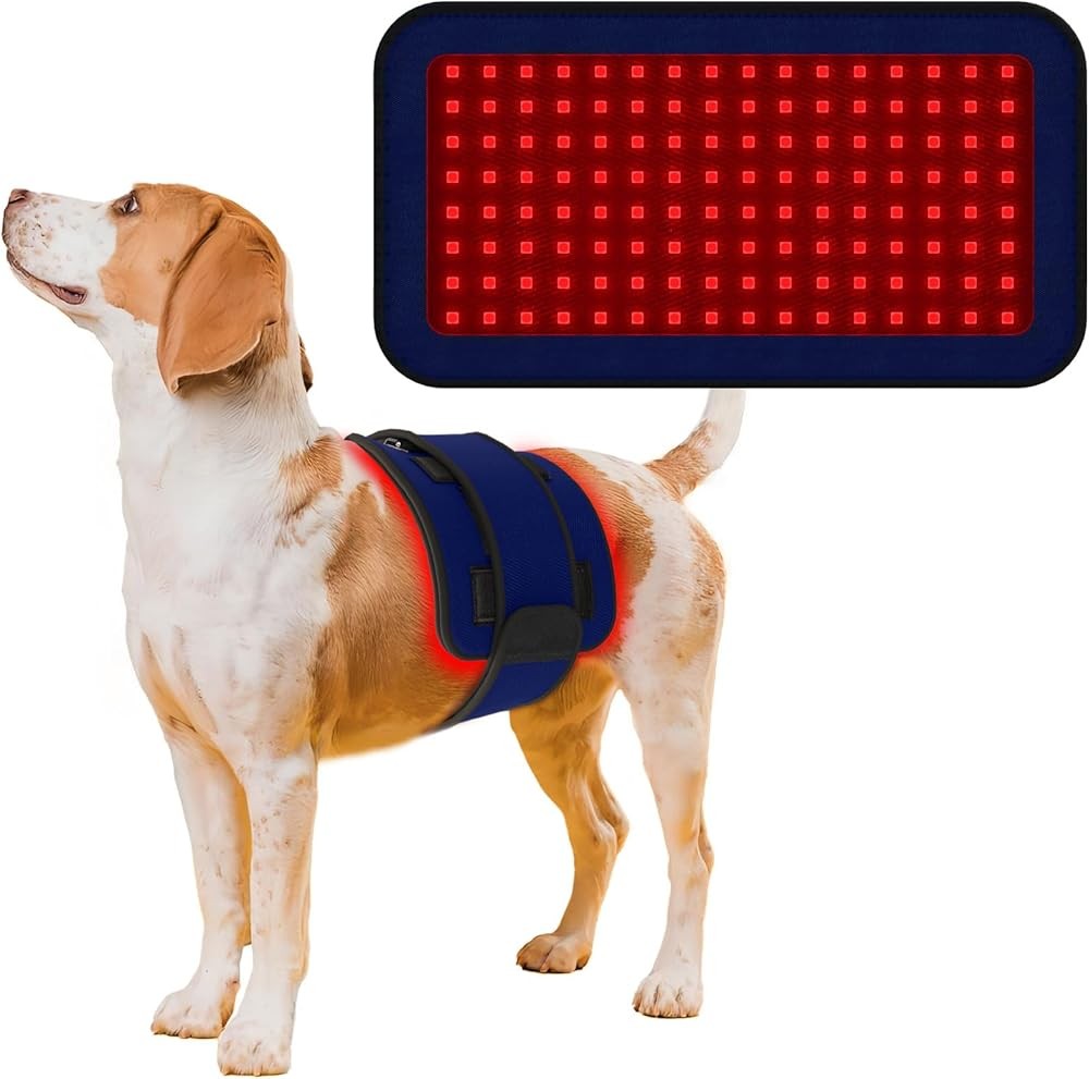
Red Infrared Light Therapy Belt for Pets
The wearable belt design wraps around the hip and lower back region, maintaining consistent contact with the treatment area throughout the session without requiring manual device holding. The belt contains 32 diodes distributed across a flexible panel that conforms to body contours, delivering combined 660nm and 850nm wavelengths.
Red Infrared Light Therapy Belt for Pets
Check Price on AmazonAs an Amazon Associate we earn from qualifying purchases.
The hands-free operation allows owners to perform other tasks during the 15-minute treatment cycle, addressing compliance challenges that arise with treatments requiring sustained attention. Dogs that resist standing still for manual device application may tolerate the belt more readily once positioned, particularly when combined with calming activities or rest periods.
The 850nm wavelength falls at the upper end of the near-infrared range, providing similar deep tissue penetration to 808nm wavelengths used in most published protocols. The 660nm red light component addresses superficial tissues, though the belt’s contact against the skin and hair coat may limit effective red light transmission compared to direct-application devices.
The belt’s wraparound design provides simultaneous coverage of bilateral hip regions and the lumbosacral area, potentially addressing not only hip dysplasia but also concurrent lower back pain that often develops secondary to altered gait mechanics in dysplastic dogs. This broader coverage area may benefit dogs with multiple pain sources more than targeted single-spot devices.
Adjustable straps accommodate dogs from approximately 30-100 pounds, though optimal fit varies by body shape and coat density. The belt should fit snugly enough to maintain diode contact with the body surface without excessive tightness that restricts movement or causes discomfort during the treatment session.
The fixed energy density of 3 J/cm² falls in the lower-moderate range of published protocols, suitable for maintenance therapy or dogs showing sensitivity to higher intensities. However, dogs requiring more aggressive protocols or having very deep tissue penetration needs may benefit from devices capable of delivering higher energy densities.
Battery operation provides portability for treating dogs in various locations or positions, with rechargeable batteries lasting approximately 10-12 sessions per charge. The built-in timer automatically shuts off after 15 minutes, ensuring consistent treatment duration and preventing device operation beyond recommended parameters.
How Does Cold Laser Therapy Combine With Other Hip Dysplasia Treatments?
Hip dysplasia management typically requires multimodal approaches addressing multiple aspects of the condition, with photobiomodulation serving as one component within a comprehensive treatment plan rather than a standalone intervention. Understanding how laser therapy interacts with other modalities helps optimize overall outcomes.
Weight management represents a fundamental intervention for hip dysplasia because excess body weight increases mechanical stress on the already compromised joint. Laser therapy addresses inflammation and pain but does not reduce the physical forces driving ongoing cartilage damage. Combining appropriate body condition maintenance with photobiomodulation provides both symptom relief and reduced disease progression.
Non-steroidal anti-inflammatory drugs (NSAIDs) remain a primary pharmaceutical intervention for hip dysplasia pain and inflammation. Laser therapy may allow reduced NSAID dosing for adequate symptom control, potentially reducing gastrointestinal and renal side effects associated with long-term NSAID use. However, laser therapy does not eliminate the need for pharmaceutical intervention in most moderate to severe cases.
Joint supplements including glucosamine, chondroitin, MSM, and omega-3 fatty acids support cartilage health and provide anti-inflammatory effects through mechanisms distinct from photobiomodulation. Published research on best dog supplements for hip and joint health reviews evidence for these nutritional interventions, which complement rather than duplicate laser therapy mechanisms.
Physical rehabilitation exercises strengthen muscles supporting the hip joint, improve range of motion, and maintain functional capacity. Combining controlled exercise protocols with pre-activity laser therapy sessions may enhance exercise tolerance and reduce post-activity soreness, though research directly testing this combination remains limited.
Specialized orthopedic dog beds for arthritis and senior dogs reduce pressure on affected joints during rest periods, complementing active laser therapy sessions. Supportive bedding does not provide therapeutic effects identical to photobiomodulation but addresses a different aspect of comprehensive comfort and joint protection.
Mobility aids including dog rehab harnesses and mobility support help dysplastic dogs maintain activity levels during treatment periods and may reduce compensatory strain on other joints. These assistive devices work synergistically with laser therapy by enabling exercise that maintains muscle mass and joint function.
Omega-3 fatty acids from sources like fish oil supplements for dogs provide anti-inflammatory effects through different biochemical pathways than photobiomodulation. The combination may produce additive or synergistic inflammation reduction, though direct studies testing laser therapy plus omega-3 supplementation are not available.
Surgical interventions including triple pelvic osteotomy (TPO), femoral head ostectomy (FHO), or total hip replacement physically alter joint anatomy to address the underlying structural problem that laser therapy cannot correct. Post-surgical laser therapy may support surgical site healing, reduce post-operative inflammation, and accelerate functional recovery, though protocols should be coordinated with the surgical veterinarian.
Intra-articular injections of platelet-rich plasma (PRP), hyaluronic acid, or corticosteroids provide localized anti-inflammatory and regenerative effects directly within the joint space. Research on combined PRP and photobiomodulation demonstrates synergistic benefits, suggesting that laser therapy enhances the efficacy of injectable joint treatments.
Acupuncture provides pain management through mechanisms involving endorphin release, nerve modulation, and circulatory effects that partially overlap with photobiomodulation mechanisms. While both modalities can be used together, the degree of mechanistic overlap suggests potentially diminishing returns rather than fully additive effects.
The evidence shows: Cold laser therapy works most effectively as part of a multimodal hip dysplasia management program combining weight management, appropriate exercise, joint supplements, pain medications as needed, and supportive care rather than as a standalone intervention replacing other evidence-based treatments.
What Are the Safety Considerations for Home Laser Therapy?
Home-use cold laser devices provide accessibility and cost advantages but require understanding safety parameters to avoid adverse effects and maximize therapeutic benefits. Class IIIb cold laser devices, the category including most home veterinary units, deliver therapeutic energy densities without causing thermal tissue damage when used according to manufacturer specifications.
Eye exposure represents the primary safety concern with laser devices because focused light energy can damage retinal structures. While therapeutic cold lasers do not produce sufficient power for immediate vision loss from brief exposure, prolonged direct viewing of the laser emission should be avoided. Safety protocols include never directing the device toward eyes, maintaining device contact with the application surface rather than pointing through air, and supervising dogs during sessions to keep them from looking directly at the light source.
Most home devices include built-in safety features that reduce eye exposure risk. Contact sensors that require skin apposition before activation stop operation while the device points at nothing, which could allow scattered light to reach eyes. Timed automatic shutoff stops extended operation beyond recommended treatment durations.
Thermal effects remain minimal with properly functioning cold lasers operating within specified parameters. Unlike Class IV high-power lasers that can cause thermal tissue damage, Class IIIb devices deliver energy below thermal damage thresholds. However, devices should not be used on a single spot for longer than recommended durations, and any sensation of heat during treatment suggests excessive energy delivery or device malfunction requiring immediate discontinuation.
Contraindications for laser therapy include application over active cancer sites, where photobiomodulation’s cellular stimulation effects could theoretically enhance tumor growth. While direct evidence of laser therapy causing cancer progression in veterinary patients remains limited, the prudent approach avoids treating areas with known malignancy until more definitive safety data exists.
Application over growth plates in young dogs requires caution because photobiomodulation affects bone cell activity. While no published evidence documents premature growth plate closure from therapeutic laser application, conservative protocols use lower energy densities and reduced session frequencies in puppies with open growth plates until skeletal maturity.
Pregnancy represents a theoretical contraindication based on unknown effects of light exposure on developing fetuses, though no actual evidence of harm exists. Avoiding laser application over the pregnant uterus follows a precautionary approach rather than responding to documented adverse outcomes.
Recent surgical sites should be evaluated before laser application to ensure adequate healing for light transmission without interference from surgical implants. Metal implants including pins, plates, and screws may cause some light scattering but do not absolutely contraindicate laser therapy. However, immediate post-operative periods may benefit from delaying laser therapy until the surgical veterinarian confirms appropriate healing.
Photosensitizing medications including certain antibiotics and anti-fungal drugs can increase tissue sensitivity to light exposure, though the specific wavelengths causing photosensitivity reactions typically differ from therapeutic near-infrared wavelengths. Reviewing current medications with a veterinarian before initiating laser therapy helps identify potential interactions.
Device maintenance affects safety and efficacy. Regular cleaning of the treatment head ensures optimal light transmission without obstruction from accumulated debris or oils. Battery-powered devices should be charged according to manufacturer specifications to avoid performance degradation or battery-related safety issues.
Here’s what matters: Class IIIb home cold laser devices provide good safety profiles when used according to manufacturer guidelines, with primary safety considerations including eye protection, avoiding application over undiagnosed masses, and using appropriate energy densities and treatment durations as prescribed by a veterinarian familiar with photobiomodulation protocols.
How Do You Monitor Your Dog’s Response to Laser Therapy?
Systematic tracking of clinical parameters helps determine whether laser therapy produces meaningful improvements, informs protocol adjustments, and identifies dogs that may not respond adequately to light therapy alone. Objective measures combined with structured observations provide more reliable assessment than casual impressions.
Mobility scoring systems provide standardized frameworks for evaluating functional changes. The Canine Brief Pain Inventory (CBPI) includes questions about pain severity and activity interference that owners complete at regular intervals, allowing quantitative comparison of pre-treatment, during-treatment, and post-treatment status. Many veterinarians familiar with osteoarthritis management can provide CBPI forms or electronic versions.
Specific functional observations include the dog’s willingness to climb stairs, jump onto furniture, rise from lying positions, and sustain activity during walks. Noting how many stairs the dog attempts, whether assistance is required for elevation changes, and how quickly the dog rises from rest provides concrete data points for tracking improvement or decline.
Gait changes observable to owners include limb use symmetry, stride length, and movement fluidity. While professional gait analysis using force plates provides precise measurements, home observations noting whether the dog consistently bears weight on the affected limb, shows equal stride length bilaterally, and moves smoothly without obvious stiffness offer practical assessment tools.
Activity level changes including play behavior, social interactions with other dogs, and sustained exercise tolerance help gauge quality of life improvements that may not be fully captured by specific mobility tests. Dogs showing improved hip comfort often spontaneously increase play, demonstrate more animated greetings, and sustain longer walks without showing post-activity soreness.
Post-activity recovery time provides useful information about functional improvement. Before treatment, note how long the dog requires to recover normal mobility after walks or play, then track whether this recovery period shortens during laser therapy. Reduced recovery time suggests improved joint tolerance for mechanical stress.
Pain-associated behaviors including restlessness at night, reluctance to lie down or stand up, protective responses when the hip area is touched, and vocalizations during movement may decrease with effective laser therapy. Documenting the frequency of these behaviors before treatment provides baseline data for comparison during therapy.
Body language cues including ear position, tail carriage, and overall posture often change when chronic pain improves. Dogs experiencing significant hip pain may show lowered tail carriage, tucked hindquarters, and generally tense body posture that relaxes as discomfort decreases. Photographs taken before and during treatment can help visualize these subtle changes.
Medication requirements provide an indirect measure of pain control. If laser therapy produces meaningful benefit, some dogs may tolerate reduced NSAID dosing while maintaining adequate comfort. Any medication changes should be implemented under veterinary guidance rather than independently by owners.
Video documentation creates objective records for comparison across time. Brief videos of the dog walking, trotting, rising from rest, and navigating stairs before treatment and at 2-week intervals during therapy allow detailed review of gait and mobility changes that may be difficult to detect through memory alone.
Response timeframes vary between individuals, with some dogs showing noticeable improvement within the first week while others require 4-6 weeks of consistent treatment before significant changes appear. Dogs showing no improvement after 8 weeks of properly administered laser therapy at appropriate parameters may be non-responders who require different treatment approaches.
Clinical insight: Systematic monitoring using standardized mobility scoring, specific functional observations, activity level changes, and documented pain behaviors provides reliable evidence for whether laser therapy produces meaningful hip dysplasia improvement in individual dogs, guiding protocol adjustments and treatment decisions.
What Breed-Specific Considerations Apply to Hip Dysplasia Laser Therapy?
Different breeds show varying hip dysplasia prevalence, severity patterns, and anatomical characteristics that influence laser therapy approaches. While fundamental photobiomodulation mechanisms remain consistent across breeds, some practical considerations optimize treatment for specific breed types.
Large and giant breeds including Labrador Retrievers, German Shepherds, Golden Retrievers, Rottweilers, and Bernese Mountain Dogs show the highest hip dysplasia prevalence, with some breeding lines approaching 70% affected. These breeds typically benefit from higher energy densities (6-8 J/cm²) due to greater tissue depth from skin surface to hip joint, requiring more photon energy to achieve therapeutic effects at target structures.
Giant breed dogs weighing over 100 pounds may require longer session times or multiple treatment spots to ensure adequate coverage of the extensive hip region. A systematic grid approach dividing each hip into 6-8 treatment zones ensures no areas receive insufficient energy density, though this extends total session time compared to smaller dogs.
Heavily muscled breeds including American Pit Bull Terriers, American Staffordshire Terriers, and Bulldogs present increased tissue density between skin surface and hip joint, potentially requiring higher energy densities or longer application times to achieve therapeutic penetration. However, the shorter bone structure in many bully breeds may partially offset increased muscle mass.
Deep-chested breeds with pronounced tuck-up including Greyhounds, Whippets, and other sighthounds often have less subcutaneous fat over the hip region, potentially allowing effective therapy at lower energy densities (4-5 J/cm²) compared to breeds with more body fat. The lean body composition brings target tissues closer to the skin surface.
Breeds with very dense or double coats including Siberian Huskies, Alaskan Malamutes, Newfoundlands, and Samoyeds may benefit from clipping hair over the treatment area to reduce light absorption and scattering by the coat. While laser therapy can be performed through normal coat, light transmission improves with hair removal, potentially enhancing therapeutic effects.
Small and medium breeds that develop hip dysplasia, including Pugs, French Bulldogs, Cocker Spaniels, and some terrier breeds, often manage well with moderate energy densities (4-6 J/cm²) and smaller treatment heads that match their anatomical scale. Oversized treatment heads designed for large-breed hips may deliver excessive coverage area for small-breed applications.
Breeds with dark skin pigmentation including some Rottweilers, Doberman Pinschers, and other breeds with black or very dark hair may show slightly reduced light penetration due to melanin absorption, though the effect is more pronounced with red wavelengths than near-infrared. Using devices with higher 808nm output or slightly extended treatment times may compensate for pigmentation effects.
Brachycephalic breeds including English Bulldogs, French Bulldogs, and Pugs often show severe hip dysplasia combined with other structural issues and may require conservative energy densities initially to assess tolerance, with gradual increases based on individual response rather than aggressive initial protocols.
Breeds developed for specific working purposes including herding breeds (Border Collies, Australian Shepherds) and sporting breeds (Labrador Retrievers, Golden Retrievers) often maintain high activity levels despite hip dysplasia, benefiting from regular laser therapy sessions that support continued function rather than limiting activity to accommodate joint disease.
Age of onset varies by breed, with some large breeds showing clinical signs as young as 6-12 months while other breeds may not develop symptomatic hip dysplasia until middle age or later. Early-onset cases may benefit from aggressive initial laser protocols to manage inflammation during critical growth periods when surgical interventions may still be viable options.
What this means: While core photobiomodulation principles apply across all breeds, optimizing energy density based on body size and tissue depth, adjusting coverage area to match anatomical scale, and considering coat density and activity level produces protocols tailored to breed-specific characteristics and hip dysplasia patterns.
How Does Cold Laser Therapy Compare to Red Light Therapy for Hip Dysplasia?
The terms “cold laser therapy” and “red light therapy” often cause confusion because they describe overlapping but not identical modalities. Understanding the technical and practical differences helps select appropriate devices and set realistic expectations for hip dysplasia management.
Cold laser therapy specifically refers to low-level laser therapy (LLLT) using coherent (laser) light sources, typically in the red to near-infrared wavelength range. The term “cold” distinguishes therapeutic lasers from surgical lasers, indicating that the device produces no thermal tissue effects. Most therapeutic lasers used in veterinary medicine deliver near-infrared wavelengths around 808-860nm from laser diodes.
Red light therapy typically describes broader LED-based devices emitting red (630-680nm) and sometimes near-infrared (780-850nm) wavelengths using light-emitting diodes rather than laser diodes. LED devices produce non-coherent light that diverges from the source rather than maintaining the focused beam characteristics of true lasers.
The coherence and collimation of laser light theoretically provides superior tissue penetration and focused energy delivery compared to LED-based red light devices. However, practical differences in therapeutic outcomes between laser and LED sources at equivalent wavelengths and energy densities remain debated, with some studies suggesting minimal clinical differences when photon dose at target tissues is matched.
For hip dysplasia specifically, the wavelength matters more than whether the source is laser or LED. Deep joint structures require near-infrared wavelengths (780-850nm) regardless of source type. Devices providing only red wavelengths (630-680nm) without near-infrared components show insufficient penetration for hip joint therapy, whether they use laser or LED technology.
Published research comparing cold laser versus red light therapy for dogs examines evidence for each modality type, finding that near-infrared wavelengths from either source show therapeutic potential for joint disease while red-only wavelengths benefit primarily superficial conditions including wounds and dermatological issues.
Panel-style red light therapy devices designed for human whole-body exposure often emit primarily red wavelengths without adequate near-infrared component for deep joint therapy. While these devices may provide some superficial benefits, they do not substitute for targeted near-infrared delivery to hip joint structures in dysplastic dogs.
True cold laser devices typically deliver higher power density than LED panels, allowing shorter treatment times to achieve equivalent energy density. A focused laser beam concentrating energy on a small treatment area delivers therapeutic doses in minutes, while broader LED panels may require longer exposure times to accumulate equivalent photon energy at target depths.
The cost difference between laser and LED devices can be substantial, with true laser devices often costing more due to laser diode technology compared to less expensive LED arrays. However, for hip dysplasia requiring deep tissue penetration, the investment in appropriate near-infrared delivery capability justifies the cost difference through superior wavelength characteristics for the intended application.
Safety considerations differ slightly between modalities, with focused laser beams requiring more stringent eye protection compared to divergent LED light. However, both device types warrant eye safety precautions during use, and the classification into Class IIIb category applies to both laser and some high-power LED devices.
Some newer devices combine laser and LED technology, using laser diodes for near-infrared wavelengths requiring deep penetration and LED sources for red wavelengths targeting superficial tissues. These hybrid approaches may offer advantages for conditions affecting multiple tissue depths, though the therapeutic benefit over single-wavelength near-infrared sources for hip dysplasia specifically remains unclear.
Research takeaway: For hip dysplasia treatment, near-infrared wavelengths (780-850nm) are essential regardless of whether delivered by laser or LED technology, making wavelength specification more important than the technical distinction between coherent laser versus non-coherent LED light sources when selecting devices.
What Does a Typical 8-Week Hip Dysplasia Laser Therapy Protocol Look Like?
Published research protocols and clinical practice guidelines provide frameworks for structuring laser therapy interventions, though individual adjustments based on response patterns optimize outcomes. A representative 8-week protocol illustrates standard intensive treatment followed by transition to maintenance.
Weeks 1-2: Initial Intensive Phase
- Session frequency: 3-4 sessions per week (every other day or 3 consecutive days followed by 1 rest day)
- Energy density: 4-6 J/cm² depending on dog size and dysplasia severity
- Treatment area: Systematic coverage of entire hip region using grid pattern with 4-6 treatment spots per hip
- Session duration: 10-15 minutes per hip for bilateral cases
- Concurrent interventions: Continue all prescribed medications, joint supplements, and activity modifications
- Response monitoring: Document baseline mobility using CBPI or similar validated scale before first session
Weeks 3-4: Continued Intensive Phase with Response Assessment
- Session frequency: Maintain 3-4 sessions weekly
- Energy density: May increase to 6-8 J/cm² if initial response is positive with no adverse effects
- Protocol adjustment: Dogs showing minimal improvement may benefit from daily sessions or higher energy density; dogs showing rapid improvement continue current protocol
- Activity modification: May cautiously increase controlled exercise if mobility shows improvement, avoiding excessive activity that could cause setback
- Response monitoring: Complete second mobility assessment at end of week 4 to document changes from baseline
Weeks 5-6: Transition Phase
- Session frequency: Begin reducing to 2-3 sessions weekly if significant improvement is documented
- Energy density: Maintain effective level from weeks 3-4
- Sustainability assessment: Evaluate whether reduced frequency maintains improvements or requires return to intensive schedule
- Integration of other modalities: Optimize concurrent supplements, weight management, and exercise protocols based on improved mobility
- Response monitoring: Track whether reduced session frequency maintains improvement or shows regression
Weeks 7-8: Early Maintenance Phase
- Session frequency: 1-2 sessions weekly if improvements are sustained
- Energy density: May reduce to 4-6 J/cm² for long-term maintenance
- Long-term planning: Determine ongoing maintenance schedule based on individual response
- Medication review: Consult with veterinarian about potential NSAID dose reduction if pain control is adequate with reduced medication plus laser therapy
- Final response assessment: Complete third mobility evaluation to document overall improvement from baseline to end of initial protocol
Post-Protocol Maintenance
- Session frequency: 1-2 weekly sessions continued indefinitely for chronic hip dysplasia
- Protocol adjustment: Increase frequency temporarily during weather changes, increased activity periods, or symptom flares
- Periodic reassessment: Formal mobility evaluation every 3-6 months to track long-term progression
- Combined approach: Maintain laser therapy as one component within comprehensive multimodal management including appropriate weight, exercise, and other interventions
Individual variation means this protocol serves as a framework requiring adjustment rather than a rigid prescription. Dogs showing minimal improvement after 4 weeks may benefit from increased session frequency, higher energy density, or investigation of whether the dysplasia is more severe than laser therapy alone can manage, potentially requiring surgical intervention.
Dogs showing rapid early improvement may transition to maintenance protocols sooner than 8 weeks, while others benefit from extended intensive phases before reducing frequency. The goal is finding the minimum effective frequency and energy density that maintains adequate symptom control while minimizing treatment burden and cumulative time commitment.
Professional veterinary laser therapy clinics often implement similar protocols with supervision and may achieve superior outcomes through professional-grade Class IV lasers delivering higher power than home devices. However, the convenience and cost savings of home therapy using properly selected Class IIIb devices makes consistent long-term treatment more accessible for many owners.
Our assessment: An 8-week initial protocol with 3-4 weekly sessions at 4-8 J/cm² energy density provides a structured approach to establishing laser therapy benefits, with transition to 1-2 weekly maintenance sessions based on individual response patterns and symptom control.
Can Laser Therapy Help Post-Surgical Hip Dysplasia Recovery?
Surgical interventions for hip dysplasia including femoral head ostectomy (FHO), triple pelvic osteotomy (TPO), and total hip replacement require significant tissue healing and rehabilitation periods where photobiomodulation may provide supportive benefits. The therapy’s mechanisms addressing inflammation, pain, and tissue repair align well with post-surgical recovery needs.
Studies examining low-level laser therapy effects on bone healing and pain signs in dogs found that laser application following orthopedic surgery accelerated bone healing, reduced post-operative pain scores, and supported earlier return to function compared to surgery alone. The research used 808nm wavelength at 4-6 J/cm² applied to surgical sites beginning 24-48 hours post-operatively.
For FHO procedures where the femoral head is surgically removed and a fibrous pseudoarthrosis develops to replace the ball-and-socket joint, laser therapy may support the formation of functional fibrous tissue and reduce inflammation during the critical healing period. However, specific FHO laser therapy research remains limited, with extrapolation from general orthopedic surgery studies providing the primary evidence base.
TPO procedures involve cutting and repositioning the pelvis to improve acetabular coverage of the femoral head, requiring bone healing at osteotomy sites. Research on photobiomodulation effects on bone shows enhanced osteoblast activity and accelerated bone remodeling, suggesting potential benefits for TPO healing, though dog-specific TPO laser therapy studies are not available.
Total hip replacement involves extensive soft tissue dissection and implant placement, creating significant post-operative inflammation and pain. Laser therapy’s anti-inflammatory and analgesic mechanisms may reduce post-operative discomfort and support rehabilitation compliance, though research specifically examining laser therapy following canine total hip replacement has not been published.
Post-surgical protocols typically delay laser therapy for 24-72 hours after surgery to allow initial wound closure and stabilization, then begin with conservative energy densities (2-4 J/cm²) applied to the surgical site and surrounding tissues. Energy density may increase gradually as healing progresses, reaching standard therapeutic levels (4-8 J/cm²) by 2-3 weeks post-operatively.
Coordination with the surgical veterinarian is essential before implementing post-surgical laser therapy because some surgeons may have specific concerns about timing, energy density, or treatment areas based on the particular surgical technique used. Metal implants present no absolute contraindication but may cause some light scattering at the implant-bone interface.
The rehabilitation period following hip surgery often includes controlled exercise protocols, therapeutic exercises, and physical therapy interventions. Combining laser therapy with these rehabilitation modalities may produce synergistic benefits, with photobiomodulation potentially enhancing exercise tolerance and reducing post-activity inflammation.
Post-surgical pain management typically involves pharmaceutical medications that may be reduced or tapered more quickly when laser therapy is included in the recovery protocol. However, any medication adjustments should be made under veterinary guidance rather than independently by owners based on perceived improvement.
Long-term outcomes following hip surgery may be enhanced by continued laser therapy during and after the acute recovery period. Even after surgical site healing is complete, ongoing photobiomodulation may support the remaining non-operated hip in dogs with unilateral surgery or provide continued anti-inflammatory effects in the operated joint as the dog ages.
The science says: Post-surgical laser therapy at conservative initial energy densities (2-4 J/cm²) progressing to standard therapeutic levels (4-8 J/cm²) shows promise for supporting bone healing, reducing inflammation, and managing post-operative pain following hip dysplasia surgery, though specific research on canine hip surgery laser protocols remains limited.
What Are the Limitations of Cold Laser Therapy for Hip Dysplasia?
Understanding what photobiomodulation cannot accomplish helps avoid unrealistic expectations and ensures appropriate integration within comprehensive hip dysplasia management rather than over-reliance on light therapy as a complete solution. Several inherent limitations affect the therapy’s role in hip dysplasia care.
Laser therapy does not correct the underlying anatomical malformation causing hip dysplasia. The shallow acetabulum, malformed femoral head, or excessive joint laxity that define dysplastic hips persist regardless of light exposure. The therapy manages symptoms including pain, inflammation, and mobility limitation but does not alter joint structure or restore normal anatomy.
Severely dysplastic hips with extensive cartilage loss, severe osteoarthritis, or complete joint destruction may respond poorly to laser therapy because the degree of tissue damage exceeds what photobiomodulation mechanisms can address. While the therapy supports cellular function in damaged tissues, it cannot regenerate completely destroyed cartilage or reverse advanced degenerative changes.
Individual response variation means some dogs show minimal or no improvement despite appropriate device selection, correct protocols, and consistent treatment adherence. Research studies documenting average improvement across treatment groups include individual dogs that do not respond, suggesting that photobiomodulation benefits some but not all dysplastic dogs through mechanisms that remain incompletely understood.
The therapy requires ongoing sessions to maintain benefits, with symptom recurrence common when treatment is discontinued. Unlike surgical interventions that produce one-time structural corrections, laser therapy provides management rather than cure, requiring long-term commitment to maintenance protocols for sustained symptom control.
Time and compliance requirements present practical limitations because protocols requiring multiple weekly sessions for extended periods demand significant owner commitment. Dogs resistant to the treatment process or owners with limited time availability may struggle with protocol adherence, reducing real-world effectiveness compared to research study conditions.
Cost considerations include both initial device purchase and opportunity cost of time spent on treatment sessions. While home devices eliminate per-session professional fees, the upfront investment of $118-199 plus years of maintenance therapy time represents a substantial commitment that may not be feasible for all owners.
Lack of standardized protocols across veterinary practice means that specific recommendations vary between practitioners, creating uncertainty about optimal parameters. The published research provides general guidance but does not establish definitive protocols for all dysplasia severity levels, dog sizes, and individual presentations.
Limited head-to-head comparative research makes evidence-based device selection difficult because most published studies use specific professional-grade devices rather than comparing outcomes between different device types, wavelengths, or energy densities. Home device selection often relies on manufacturer claims rather than independent comparative effectiveness data.
The therapy cannot replace weight management, appropriate exercise, or other fundamental interventions that address mechanical stress on dysplastic joints. Laser therapy on an obese dog with uncontrolled activity produces inferior outcomes compared to combining photobiomodulation with optimal body condition and appropriate exercise modification.
Contraindications and precautions limit application in some cases, including dogs with concurrent cancer where cellular stimulation effects raise theoretical concerns, very young puppies with open growth plates where conservative approaches are warranted, and pregnant dogs where fetal effects remain unknown.
Here’s what matters: Cold laser therapy serves as a supportive management tool within multimodal hip dysplasia care but cannot correct underlying anatomical problems, may not benefit all individual dogs, requires ongoing maintenance for sustained effects, and works most effectively when combined with weight management, appropriate exercise, and other evidence-based interventions.
Related Reading
For comprehensive information on therapeutic cold laser applications beyond hip dysplasia, see our guide on the best cold laser therapy device for dogs covering device selection criteria, wavelength specifications, and applications across multiple conditions.
Arthritis management using photobiomodulation extends beyond hip joints to elbows, stifles, and spinal regions, as detailed in our article on cold laser therapy for dog arthritis reviewing protocols for different joint locations and arthritis types.
Wound healing represents another major application for light therapy, with specific protocols differing from joint disease approaches as explained in our guide to cold laser therapy for dog wound healing covering tissue repair mechanisms and wound-specific parameters.
Home implementation of professional-grade protocols requires understanding device capabilities and treatment techniques covered in our article on veterinary laser therapy at home comparing home devices to professional Class IV systems.
Technical differences between modalities and appropriate applications are clarified in our comparison of cold laser versus red light therapy for dogs examining wavelength effects, penetration depths, and condition-specific recommendations.
Nutritional support for hip and joint health complements physical therapies as reviewed in our guide to best dog supplements for hip and joint health covering glucosamine, chondroitin, MSM, and other evidence-based ingredients.
Supportive care during rest periods reduces joint stress through appropriate bedding as detailed in our article on best orthopedic dog beds for arthritis and senior dogs examining memory foam, gel, and other support technologies.
Mobility assistance during recovery or advanced dysplasia stages helps maintain function as explained in our guide to best dog rehab harness mobility support covering different harness types and fitting considerations.
Anti-inflammatory support through omega-3 fatty acids complements other hip dysplasia interventions as reviewed in our article on best fish oil supplements for dogs examining EPA and DHA content and bioavailability factors.
Recommended Products
Get Weekly Research Updates
New studies, updated reviews, and evidence-based health insights delivered to your inbox. Unsubscribe anytime.