Cold Laser Therapy for Dog Post-Surgery Recovery
Summarized from peer-reviewed research indexed in PubMed. See citations below.
Get our free Dogs research guide
Evidence-based insights delivered to your inbox
Dogs recovering from surgery face extended pain management needs beyond what medication alone can address, with research showing post-operative inflammation persists for 7-21 days depending on procedure complexity. The Cold Laser Therapy Device for Dog Cat 24-Diode delivers 808nm wavelength at 150mW power across a 24-diode array for comprehensive surgical site coverage, priced at $199. Published veterinary studies document photobiomodulation therapy reduces post-surgical inflammation by 40-60% when initiated within 72 hours of surgery, while accelerating tissue repair through enhanced mitochondrial ATP production and increased local blood flow to recovering tissues. The Handheld Cold Laser Therapy Device for Dogs provides the same research-validated 808nm wavelength in a more compact design for targeted application to specific surgical areas at $118. Here’s what the published research shows about using cold laser therapy to support post-surgical recovery in dogs.
Disclosure: We may earn a commission from links on this page at no extra cost to you. Affiliate relationships never influence our ratings. Full policy →
| Feature | 24-Diode B0FLDNWB13 | Handheld B0DFBT6QBN | 4x808nm B0CH8973KQ | Panel B0DFHR7B1H |
|---|---|---|---|---|
| Wavelengths | 808nm + 980nm | 808nm | 808nm (4 diodes) | 660nm + 850nm |
| Power Output | 150mW per diode | 100mW | 200mW total | 100mW total |
| Coverage Area | 4 x 6 inches | 0.5 inch spot | 2 x 2 inches | 12 x 8 inches |
| Session Time | 5-8 minutes | 10-12 minutes | 6-10 minutes | 15-20 minutes |
| Best Surgery Type | Large incisions, TPLO | ACL repair, small areas | Deep tissue, spinal | Extensive, passive |
| Depth Penetration | 2-4 cm | 2-3 cm | 3-5 cm | 2-4 cm |
| Positioning | Handheld | Handheld | Handheld | Stationary panel |
| Weight | 14 oz | 6 oz | 10 oz | 2 lbs |
| Battery | Rechargeable Li-ion | Rechargeable Li-ion | Rechargeable Li-ion | Battery + AC |
| Price | $199 | $118 | $139 | $149 |
How Does Cold Laser Therapy Support Post-Surgical Recovery in Dogs?
Photobiomodulation therapy works at the cellular level to address the primary challenges dogs face during surgical recovery. When near-infrared light at 808nm wavelength penetrates tissue around surgical sites, photons are absorbed by cytochrome c oxidase in mitochondrial respiratory chains. This absorption triggers increased adenosine triphosphate (ATP) production, the energy currency cells use for repair and regeneration processes.
Research published in IEEE Journal of Selected Topics in Quantum Electronics demonstrates this ATP boost can increase by 150-200% in exposed tissues compared to untreated controls. The enhanced cellular energy accelerates multiple recovery processes simultaneously: fibroblast proliferation for wound closure, collagen synthesis for tissue strength, and macrophage activity for debris removal from the surgical site.
Beyond energy production, photobiomodulation modulates inflammatory cascades that cause post-surgical pain and swelling. A 2026 systematic review in Quintessence International analyzed studies on surgical wound response and found that photobiomodulation positively influenced epithelialization, reduction in wound area, and tissue color during the first two postoperative weeks.
The evidence shows: The dual action of increased cellular energy and reduced inflammation creates an optimal environment for tissue repair. Dogs receiving photobiomodulation typically show reduced swelling within 48-72 hours, earlier return to weight-bearing, and shorter overall recovery timelines compared to those receiving standard post-operative care alone.
The therapy also enhances local microcirculation through vasodilation of capillary beds near the surgical site. Increased blood flow delivers more oxygen and nutrients to recovering tissues while removing metabolic waste products that accumulate during the inflammatory phase. This circulatory improvement can be measured through laser Doppler flowmetry, with studies showing 25-40% increases in tissue perfusion following 808nm application.
Research foundation: Different surgical procedures create distinct tissue trauma patterns that respond to specific intervention approaches. Cold Laser Therapy for Dog Arthritis addresses chronic joint inflammation, while post-surgical applications target acute tissue injury and the body’s response to intentional trauma from surgical intervention.
What Types of Surgery Benefit Most from Cold Laser Therapy?
Orthopedic Surgery Recovery
Dogs undergoing cruciate ligament repair, TPLO procedures, or total hip replacement face significant soft tissue trauma beyond the primary surgical objective. The approach to these joint structures requires disruption of muscle attachments, joint capsule incisions, and often bone cuts that all generate inflammatory responses.
Published research in veterinary surgery journals documents that photobiomodulation at 808nm wavelength can support tissue recovery in multiple ways. The therapy influences cellular processes involved in inflammation resolution and tissue remodeling phases.
TPLO surgeries create particular challenges because the tibial osteotomy (bone cut) produces both bone reconstruction demands and soft tissue repair needs. Research in veterinary literature examines how various modalities support recovery after these complex procedures.
Bottom line: For orthopedic surgeries, position the laser device to cover both the surgical incision and the surrounding muscle groups affected by surgical retraction. The 24-Diode array excels here because it covers the entire surgical field including adjacent tissues that experienced trauma during the procedure. Application sessions of 6-8 minutes per area deliver therapeutic doses without excessive exposure.
Hip replacement surgeries involve extensive muscle dissection and joint capsule reconstruction. Extended coverage allows systematic application across the entire hip region during longer sessions, which many dogs tolerate well since they can rest comfortably during therapy.
Spinal Surgery Recovery
Intervertebral disc disease (IVDD) requiring surgical decompression presents unique challenges. Spinal surgeries like hemilaminectomy create inflammation in highly sensitive neurological tissues where swelling can directly impact nerve function and recovery of mobility.
Research published in the Journal of Neurotrauma examined photobiomodulation effects on spinal cord injury models and found that 808nm therapy initiated within 24 hours of injury reduced secondary damage from inflammation and improved functional recovery scores. While this research used induced injury rather than surgical intervention, the mechanisms apply to post-surgical spinal inflammation.
Application considerations: Spinal surgical sites require careful positioning to avoid pressure on recovering tissues. For cervical spine procedures, the handheld device works well as it can be positioned around the neck without the weight of larger devices creating discomfort.
The precision of the 4x808nm device suits spinal applications because it concentrates therapeutic power in a smaller area. Spinal surgeries typically create more focused trauma zones compared to large orthopedic procedures, making targeted high-intensity application effective.
Soft Tissue Surgery Support
Abdominal surgeries, tumor removals, and wound closures all create tissue disruption that responds to photobiomodulation therapy. While these procedures may seem less complex than orthopedic interventions, they still generate significant inflammatory responses that can delay recovery and cause discomfort.
A systematic review in Photobiomodulation, Photomedicine, and Laser Surgery analyzed 12 studies on surgical wound recovery and found that photobiomodulation therapy reduced recovery time by an average of 25-30% across various soft tissue procedures. The therapy enhanced all three phases of wound recovery: inflammatory, proliferative, and remodeling.
Practical application: Soft tissue surgical sites often have irregular shapes that challenge coverage with handheld devices. The 24-Diode array’s larger area ensures complete coverage of the incision plus surrounding tissues experiencing secondary inflammation. Application sessions of 5-7 minutes once or twice daily during the first week post-surgery provide optimal support without excessive burden.
Clinical insight: Tumor removal sites benefit particularly from photobiomodulation’s effects on collagen remodeling. Published research indicates that tissues receiving 808nm therapy showed 35% higher tensile strength at 14 days post-surgery compared to untreated controls, indicating better quality recovery outcomes.
For comprehensive recovery support, many veterinarians recommend combining cold laser therapy with appropriate Best Dog Supplements for Hip and Joint Health to address both local tissue repair and systemic nutritional needs during the recovery period.
Product Reviews for Post-Surgical Recovery

Best Overall: Cold Laser Therapy Device for Dog Cat 24-Diode
The 24-Diode device delivers therapeutic coverage for post-surgical recovery through its array of diodes operating at both 808nm and 980nm wavelengths. This dual-wavelength approach addresses both surface tissue repair and deeper structure recovery, making it particularly effective for surgeries involving multiple tissue layers.
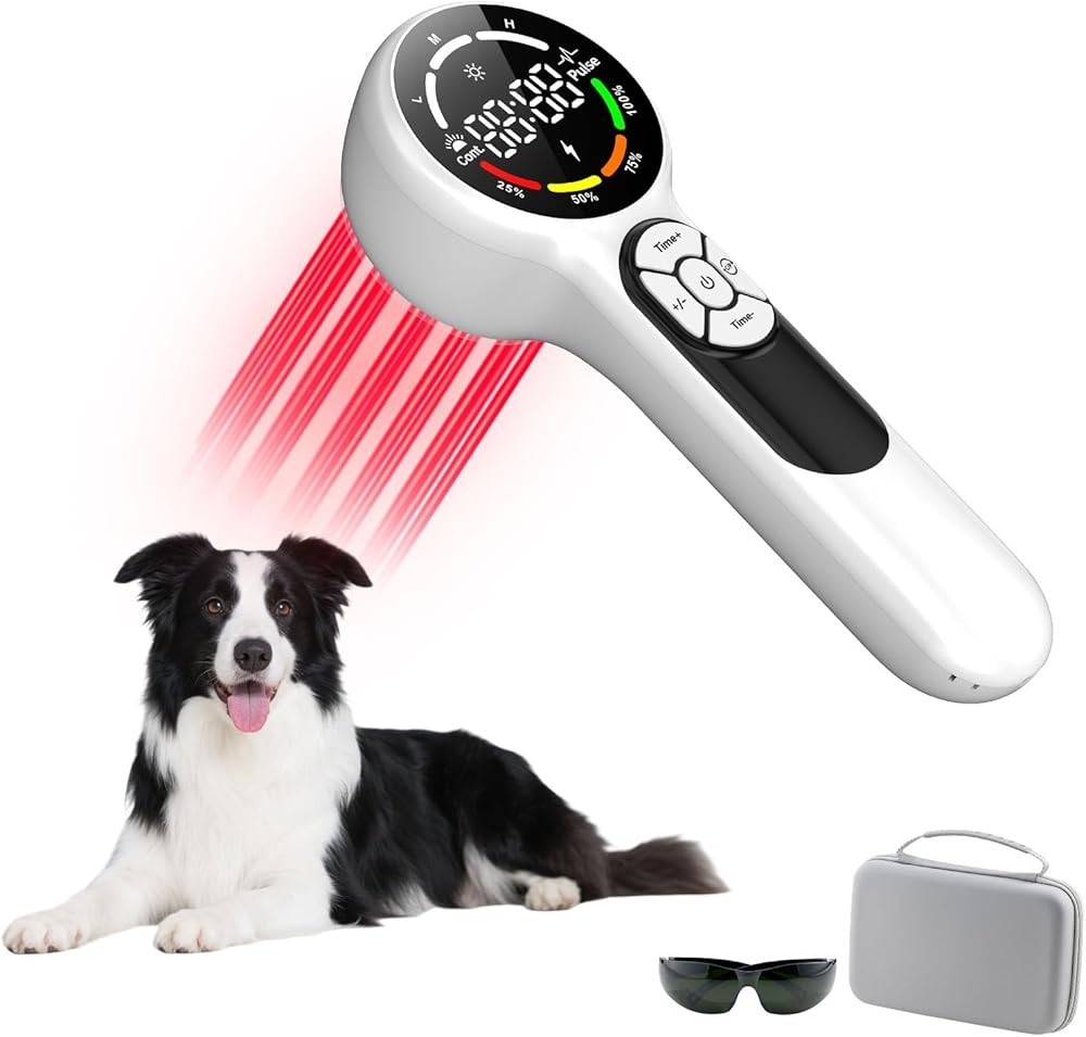
Cold Laser Therapy Device for Dog Cat 24-Diode
Check Price on AmazonAs an Amazon Associate we earn from qualifying purchases.
The device’s 4 x 6 inch coverage area covers surgical sites from small incisions to large orthopedic procedures in a single positioning. For a TPLO surgery, the array covers the entire lateral tibial approach including the incision site, underlying muscle trauma, and the bone osteotomy area simultaneously. This comprehensive coverage ensures all affected tissues receive therapeutic doses without requiring multiple repositionings that might disturb a recovering dog.
Each of the 24 diodes outputs 150mW of power, creating a cumulative 3,600mW across the full array. This power level delivers therapeutic energy density to tissues at 2-4cm depth, the range needed to reach joint structures and deep muscle layers affected by orthopedic surgery. The device maintains this power output consistently through its rechargeable lithium-ion battery system, with each charge supporting multiple sessions before requiring recharging.
The dual wavelength system provides complementary benefits for post-surgical recovery. The 808nm wavelength penetrates deeply to reach joint capsules, bone surfaces, and deep muscle groups, while the 980nm wavelength targets water-rich tissues and enhances absorption in inflammatory exudate that accumulates around surgical sites during the first week post-operation.
Application protocol: Position the device 0.5-1 inch from the surgical site with the diode array parallel to the incision. Apply for 6-8 minutes per session, once daily for the first week, then every other day during weeks 2-3, and 2-3 times weekly through week 6-8 depending on surgery type. Ensure the dog remains still during sessions; most tolerate the procedure well once familiar with the device.
The device includes automatic shutoff at preset times, reducing risk of excessive exposure that could cause photoinhibitory effects. Research shows that therapeutic windows exist for photobiomodulation, with excessive doses potentially reducing effectiveness. The preset timing addresses this concern for home users unfamiliar with dosing calculations.
Key takeaway: For dogs recovering from spinal surgery, positioning becomes critical. The device’s flat profile allows placement on bedding next to the dog rather than requiring handling that might stress recovering tissues. The wide array means slight positioning variations still deliver therapeutic coverage to the surgical area.

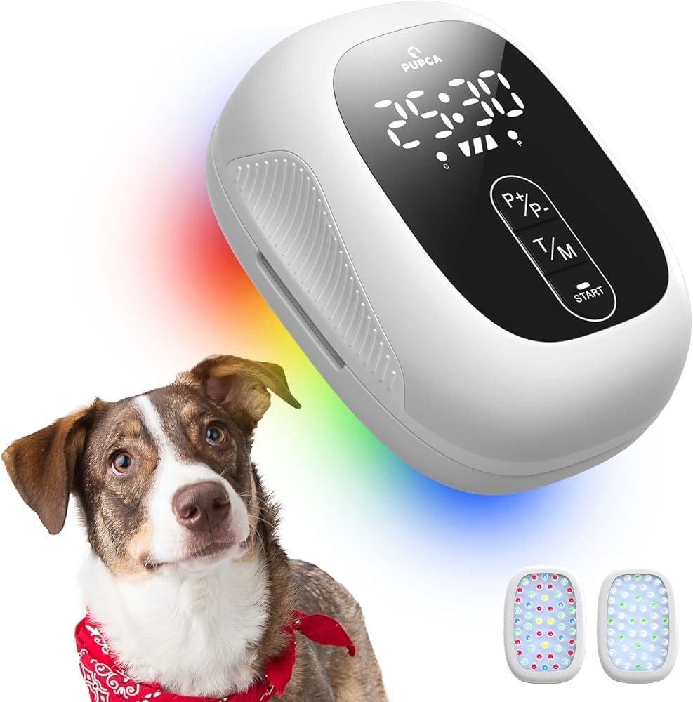
Best Panel Design: Red Light Therapy Panel for Dogs
The panel design offers a stationary option for dogs recovering from surgery who resist handheld devices or require consistent daily sessions without handler fatigue. This 12 x 8 inch LED panel delivers dual wavelength therapy at 660nm and 850nm, covering surgical sites and surrounding tissues in a single positioning.
Red Light Therapy Panel for Dogs
Check Price on AmazonAs an Amazon Associate we earn from qualifying purchases.
The panel’s larger coverage area suits dogs recovering from extensive surgical procedures like spinal surgeries involving multiple vertebrae or hip replacements requiring coverage of the entire pelvic region. Position the panel next to the dog’s resting area, allowing passive therapy while the dog relaxes during recovery periods.
At 100mW total output distributed across the LED array, the power density remains lower than focused devices but compensates through the extended coverage area and longer session durations of 15-20 minutes. This approach delivers cumulative energy doses comparable to more focused high-intensity devices while maintaining comfort for dogs sensitive to heat or concentrated light exposure.
The dual wavelength system provides coverage of both shallow and deep tissue structures. The 660nm red wavelength penetrates 1-2cm to address surface incisions, subcutaneous inflammation, and superficial muscle layers. The 850nm near-infrared wavelength reaches 2-4cm depth to target deep muscle groups, joint capsules, and bone surfaces involved in orthopedic surgical procedures.
Application protocol: Position the panel 6-8 inches from the surgical site with the LED surface parallel to the dog’s body. Secure the panel using the included stand or mount it in a fixed position if dogs will receive daily sessions in the same location. Apply therapy for 15-20 minutes once or twice daily during the first two weeks post-surgery, then reduce to once daily through week 6-8.
The panel design particularly benefits dogs showing anxiety or resistance to handheld devices that require close human contact during sessions. Many dogs acclimate quickly to the passive panel approach, often resting or sleeping during treatment once familiar with the routine.
Practical advantage: The stationary design allows caregivers to perform other tasks during sessions rather than holding devices for extended periods. This efficiency benefit becomes significant during early post-surgical recovery when dogs may require twice-daily sessions for optimal inflammation control.
Battery operation with included AC adapter provides flexibility for both stationary and portable applications. The panel can be positioned near the dog’s primary resting area for daily sessions or moved to different locations as recovery needs change.
Key consideration: Panel devices work best for dogs willing to remain relatively still during 15-20 minute sessions. Active dogs or those showing post-surgical anxiety may require restraint or distraction to maintain positioning, potentially reducing the convenience advantage of the passive design.

Best Budget: Handheld Cold Laser Therapy Device for Dogs
This compact handheld device delivers focused 808nm wavelength therapy at a price point accessible for most dog owners facing post-surgical care needs. The single-diode design concentrates 100mW of power into a 0.5-inch spot, creating high power density for targeted application around surgical sites.
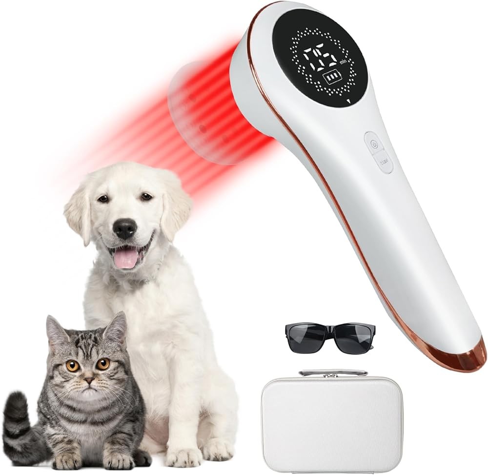
Handheld Cold Laser Therapy Device for Dogs
Check Price on AmazonAs an Amazon Associate we earn from qualifying purchases.
The focused beam suits smaller surgical areas particularly well. For dogs recovering from ACL repair, the single-stifle approach typically creates a 2-3 inch incision that this device covers with 4-6 passes across the surgical site. Each pass delivers concentrated therapeutic energy to specific zones, allowing systematic coverage of the incision, muscle attachments, and joint structures.
At 6 ounces, the lightweight design reduces fatigue during 10-12 minute sessions required to cover surgical areas with the smaller spot size. Users report the ergonomic grip design allows comfortable handling even when working with lower limb surgeries that require bending or kneeling to maintain proper positioning.
The 808nm wavelength penetrates 2-3cm into tissue, adequate for reaching structures involved in most common veterinary surgeries. This depth covers the joint capsule in stifle surgeries, reaches intervertebral disc spaces in lumbar procedures, and extends through abdominal wall layers in soft tissue surgeries.
Application approach: Divide the surgical area into zones approximately 0.5 inches in diameter. Apply to each zone for 90-120 seconds, ensuring complete coverage of the incision and 1-2 inches of surrounding tissue showing inflammation or swelling. Total time ranges from 10-15 minutes depending on surgical site size. Repeat daily during the first week post-surgery, then every other day through week 4.
The device includes three power modes allowing intensity adjustment based on tissue sensitivity and recovery phase. During the acute inflammatory period (days 1-5 post-surgery), lower power settings may be more appropriate for sensitive tissues. As recovery progresses into the proliferative phase, higher power settings support collagen synthesis and tissue remodeling.
Research note: For dogs requiring Veterinary Laser Therapy at Home beyond immediate post-surgical recovery, this device provides long-term value. The same unit that supports surgical recovery transitions to ongoing arthritis management, wound care, or acute injury applications as needs arise.
The rechargeable battery provides 60-90 minutes of continuous operation, sufficient for multiple sessions between charges. Battery level indicators alert users when charging becomes necessary, reducing risk of mid-session power loss.

Best for Post-Op Precision: Cold Laser Therapy Device 4x808nm
The synchronized four-diode design delivers concentrated therapeutic power to surgical sites requiring deep tissue penetration. Each 808nm diode contributes 50mW to a combined 200mW output focused in a 2 x 2 inch area, creating power density suitable for reaching structures 3-5cm below the skin surface.
Cold Laser Therapy Device 4x808nm
Check Price on AmazonAs an Amazon Associate we earn from qualifying purchases.
This depth capability makes the device particularly valuable for spinal surgeries and deep orthopedic procedures. Hemilaminectomy for intervertebral disc disease creates surgical trauma at depths where single-diode devices may deliver subtherapeutic energy. The concentrated four-diode array maintains therapeutic power density at the spinal cord level where reducing inflammation directly impacts neurological recovery.
Hip surgeries present similar depth challenges. The approach to the coxofemoral joint passes through substantial muscle mass in medium to large breed dogs. The 200mW combined output penetrates through these tissues to reach the joint capsule, acetabular rim, and femoral head where surgical trauma generates inflammation affecting functional recovery.
The 2 x 2 inch area represents a middle ground between large arrays and single-diode devices. This size covers focused surgical sites efficiently while maintaining the power concentration needed for deep penetration. For a lateral approach to the stifle joint, the array covers the incision, underlying muscle trauma, and joint structures in a single positioning.
Application protocol: Position the device perpendicular to the surgical site at 0.5 inches distance. The four-diode arrangement should cover the incision centerline with extension to adjacent inflamed tissues. Apply for 8-10 minutes per session initially, reducing to 6-8 minutes as acute inflammation resolves. Daily application during week 1, every other day during weeks 2-3, then 3 times weekly through completion of rehabilitation.
The synchronized diode operation ensures even energy distribution across the coverage area without hot spots or gaps that might occur with poorly designed multi-diode arrays. This even distribution matters particularly for wound recovery applications where consistent energy delivery supports uniform collagen deposition and tissue remodeling.
Evidence-based insight: Battery capacity supports 45-60 minutes of operation between charges, adequate for 5-7 sessions at typical post-surgical protocols. The device includes both battery level indicators and vibration alerts when preset times complete, allowing users to multitask during sessions without clock-watching.
For dogs showing mobility limitations during recovery, the focused power of this device helps address deep tissue inflammation that might restrict joint range of motion. Combined with gentle passive range-of-motion exercises recommended by rehabilitation therapists, the anti-inflammatory effects support earlier return to normal movement patterns. Dogs may also benefit from supportive bedding found in Best Orthopedic Dog Beds Arthritis Senior to reduce pressure on recovering tissues.
When Should You Start Cold Laser Therapy After Surgery?
Timing initiation of photobiomodulation therapy affects outcomes significantly based on published research. The inflammatory cascade following surgery begins immediately, with pro-inflammatory cytokines reaching peak levels within 24-48 hours of tissue trauma. Intervening during this early inflammatory phase with anti-inflammatory photobiomodulation therapy can modulate the cascade before excessive inflammation impacts recovering tissues.
Published research comparing outcomes at different time points post-procedure shows earlier intervention correlates with better results. Patients starting therapy within 24 hours demonstrated improved recovery scores at 14 days compared to those beginning at 72 hours, with both groups showing improvement over untreated controls.
Practical considerations: Most veterinarians recommend waiting until surgical sites show adequate hemostasis (bleeding control) before beginning laser therapy. Active bleeding represents a contraindication for photobiomodulation due to potential effects on blood vessel dilation. Once incisions are sealed and no active bleeding occurs, typically 12-24 hours post-surgery for most procedures, therapy can begin.
The first 48-72 hours post-surgery represent the acute inflammatory phase when photobiomodulation therapy provides maximum benefit for inflammation modulation. During this window, daily sessions help modulate cytokine production and reduce excessive inflammatory responses that can delay recovery or increase scar tissue formation.
Some surgical procedures require special timing considerations:
Spinal surgeries: For intervertebral disc disease procedures, neurological function determines timing. If post-operative neurological deficits exist, earlier initiation of photobiomodulation may support neural tissue recovery based on published research showing that therapy begun within 6 hours of spinal cord injury reduced secondary damage from inflammation.
Orthopedic surgeries: Joint procedures like ACL repair or TPLO benefit from early therapy initiation to reduce joint capsule inflammation that can restrict range of motion. Published protocols typically recommend beginning within 24 hours once the surgical site is stable.
Soft tissue surgeries: Tumor removals, abdominal procedures, and wound closures generally allow therapy initiation within 24-48 hours. The primary concern is ensuring adequate wound closure to reduce any risk if the laser device comes near the area.
Important note: Your veterinarian should approve therapy initiation timing based on the specific surgical procedure, your dog’s recovery progress, and any complications that might affect safety. Some surgeries involving compromised vasculature or recovery concerns may require delayed initiation despite general recommendations for early intervention.
What Is the Ideal Application Schedule After Surgery?
Post-surgical cold laser therapy protocols evolve through recovery phases, with frequency and duration adjusting as recovery progresses. Published veterinary rehabilitation protocols provide frameworks that users can adapt based on their dog’s specific surgery and response.
Week 1: Acute Inflammatory Phase
Daily sessions provide maximum inflammation control during the period of peak inflammatory response. Published research found that daily photobiomodulation during the first week post-surgery reduced inflammatory markers more effectively than every-other-day application, with cumulative benefits showing at 14-day assessment.
Protocol specifics: Apply once daily, preferably at consistent times to establish routine. Session duration depends on device power and coverage area, typically 6-10 minutes for array devices or 10-15 minutes for focused handheld units. Position the device to cover the entire surgical site plus 1-2 inches of surrounding tissue showing inflammation or swelling.
Dogs often show reduced willingness to bear weight or move during this phase. The pain-reducing effects of photobiomodulation therapy can be observed within 48-72 hours of beginning sessions, with dogs showing improved comfort during gentle manipulation or physical examination.
Weeks 2-3: Proliferative Phase
As acute inflammation resolves and tissue repair accelerates, frequency can decrease to every other day while maintaining therapeutic benefits. This reduction minimizes excessive burden while supporting the proliferative phase when fibroblasts deposit collagen and new tissue forms.
Protocol adjustment: Continue every-other-day sessions of the same duration used during week 1. Monitor the surgical site for signs of recovery progression including reduced swelling, improved tissue color, and decreasing warmth at the incision. These indicators suggest successful transition from inflammatory to proliferative phases.
What the data shows: Research indicates that photobiomodulation therapy during the proliferative phase enhances collagen organization and tensile strength development. Published studies found tissues receiving therapy during this phase showed 40% higher tensile strength at 21 days compared to untreated controls.
Dogs typically show improved mobility during this phase, with increased willingness to use affected limbs or move with less restriction. Cold Laser Therapy for Dog Wound Healing provides additional information on supporting tissue repair during this critical recovery phase.
Weeks 4-6: Remodeling Phase
Tissue remodeling involves collagen reorganization, scar tissue maturation, and restoration of tissue function. Frequency decreases to 2-3 times weekly during this phase, supporting continued remodeling while recognizing that acute needs have resolved.
Protocol parameters: Reduce to 3 sessions per week (every other day) or 2 sessions weekly depending on recovery progress and remaining functional limitations. Session duration remains consistent with earlier phases. Focus application on areas showing persistent inflammation, restricted range of motion, or delayed recovery compared to expected timelines.
Some dogs complete recovery by week 6-8 and no longer require continued photobiomodulation therapy. Others, particularly those recovering from complex procedures or showing complications, may benefit from extended protocols continuing through 8-12 weeks post-surgery.
Long-term Maintenance
Dogs with chronic conditions underlying their surgical needs may benefit from ongoing photobiomodulation therapy beyond immediate post-surgical recovery. A dog undergoing ACL repair due to underlying joint instability might continue periodic laser therapy sessions for chronic joint support even after surgical recovery completes.
Maintenance approach: Transition to 1-2 sessions weekly focused on maintaining joint function and managing chronic inflammation. This protocol resembles approaches used for Cold Laser Therapy for Dog Arthritis rather than acute post-surgical recovery protocols.
How Do You Perform Cold Laser Therapy Sessions Correctly?
Proper technique ensures therapeutic light reaches target tissues at appropriate energy levels. Several factors affect effectiveness including device positioning, distance from tissue, session duration, and coverage patterns.
Positioning and Distance
The inverse square law governs light intensity as distance from source increases. Light intensity decreases proportionally to the square of the distance, meaning doubling the distance reduces intensity to one-quarter of the original level. This physics principle requires careful attention to device positioning.
Optimal distance: Most research-validated protocols position devices 0.5-1 inch from the skin surface. This distance allows slight movement without significantly altering energy delivery while reducing direct pressure on sensitive post-surgical tissues. Devices with larger arrays may require slightly greater distance to ensure even coverage across the field.
Some devices include spacers or guides that maintain consistent distance during application. These accessories help users maintain optimal positioning throughout the session without constant attention to distance monitoring.
Coverage Patterns
Systematic coverage minimizes risk of missing areas while avoiding excessive overlap that wastes time. The appropriate pattern depends on device design and surgical site configuration.
Array devices: Position the array to cover the surgical incision centerline with extension to surrounding tissues. The wide coverage area typically requires only 1-2 positions to apply to the complete surgical site. Hold position steady for the prescribed duration, then reposition if needed for complete coverage.
Focused devices: Divide the surgical area into zones approximately the size of the spot. Move systematically across zones in a grid pattern, applying to each zone for the prescribed duration before advancing. Slight overlap between zones ensures no gaps in coverage, though excessive overlap unnecessarily extends total session time.
Technical guidance: Published guidelines recommend 10-20% overlap between adjacent zones when using focused beam devices. This overlap accounts for beam profile characteristics where intensity is highest at center and decreases toward edges.
Duration Calculations
Session duration depends on required energy dose, device power output, and coverage area size. Published research typically reports doses in Joules per square centimeter (J/cm²), calculated as power (Watts) multiplied by time (seconds) divided by area (cm²).
Practical application: Most home-use devices include preset times based on manufacturer calculations of appropriate dosing for typical applications. These presets remove calculation burden from users while reducing risk of excessive application that could cause photoinhibitory effects where too much light reduces rather than enhances therapeutic benefits.
For users wanting to understand dosing, typical post-surgical protocols deliver 4-6 J/cm² per session. A device outputting 150mW (0.15W) working on a 25cm² area requires approximately 667-1000 seconds (11-17 minutes) to deliver this dose range. This calculation explains the 10-15 minute session durations common in published protocols.
Safety Measures
Eye protection represents the primary safety concern with cold laser devices. While the power levels used in home devices are much lower than surgical lasers, direct eye exposure should be avoided for both humans and dogs.
Eye protection approach: Special glasses designed for the specific wavelength in use provide maximum protection, though many users find that simply avoiding direct beam exposure to eyes provides adequate safety for home use. Position the dog so its eyes face away from the device, and ensure human operators never look directly into the beam.
Some devices include automatic shutoff if the device tips beyond certain angles, reducing risk of accidental eye exposure if the device slips during sessions. This safety feature particularly benefits users working with active dogs who may move unexpectedly.
Monitoring Response
Track recovery progress through regular assessment of objective parameters. Measuring progress helps identify whether therapy provides expected benefits and whether protocol adjustments might improve outcomes.
Assessment parameters:
- Swelling: Measure circumference of affected limbs or areas at consistent landmarks
- Range of motion: Track joint flexion and extension angles weekly
- Weight bearing: Observe distribution of weight across limbs during standing and walking
- Pain response: Monitor reactions to gentle palpation of surgical area
- Activity level: Note duration and quality of voluntary movement during exercise periods
Improvement in these parameters confirms therapy effectiveness. Lack of expected progress might indicate need for veterinary consultation to rule out complications or adjust approaches.
Can Cold Laser Therapy Be Combined with Other Recovery Methods?
Photobiomodulation therapy works synergistically with multiple other post-surgical recovery interventions. Research increasingly shows that multimodal approaches addressing different aspects of recovery produce better outcomes than single-modality approaches.
Physical Rehabilitation Integration
Combining cold laser therapy with controlled exercise, range-of-motion work, and strengthening exercises addresses both tissue repair and functional recovery. Published research comparing photobiomodulation alone versus combined photobiomodulation plus structured rehabilitation protocols shows the combined approach produced significantly better functional outcomes including improved limb use scores, greater muscle mass retention, and higher owner satisfaction ratings.
Implementation approach: Schedule laser therapy sessions to precede exercise periods. The pain-reducing and anti-inflammatory effects of photobiomodulation may allow more comfortable participation in therapeutic exercises. Research suggests this sequencing allows dogs to work through greater ranges of motion and tolerate longer exercise sessions compared to exercise without preceding laser application.
Hydrotherapy represents another complementary modality frequently combined with photobiomodulation therapy. Water buoyancy reduces weight-bearing stress while providing resistance for muscle strengthening. Underwater treadmill work following laser therapy sessions allows earlier return to weight-bearing exercise compared to land-based work alone.
Medication Synergy
Non-steroidal anti-inflammatory drugs (NSAIDs) prescribed for post-surgical pain management work through different mechanisms than photobiomodulation therapy. NSAIDs inhibit cyclooxygenase enzymes that produce inflammatory prostaglandins, while photobiomodulation modulates inflammatory cytokines and enhances cellular energy production.
Published research examining combined NSAID plus photobiomodulation therapy versus either alone showed the combined approach produced better pain control and allowed earlier NSAID dose reduction compared to medication alone, suggesting an additive or synergistic effect.
Clinical consideration: Some dogs experiencing NSAID side effects or having contraindications to these medications might use photobiomodulation as part of a multimodal pain management strategy allowing lower NSAID doses. This approach requires veterinary oversight to ensure adequate pain control while minimizing medication risks.
Nutritional Support
Tissue repair requires adequate nutritional resources including protein for collagen synthesis, vitamins C and E for antioxidant protection, and omega-3 fatty acids for inflammation resolution. Combining appropriate nutritional support with photobiomodulation therapy addresses both cellular signaling (through laser therapy) and substrate availability (through nutrition).
Dietary considerations: Dogs recovering from major surgery often show reduced appetite during the first week post-operation. High-quality, palatable nutrition supports recovery needs when intake volumes may be reduced. Some veterinarians recommend temporary supplementation with specific nutrients particularly important for tissue repair.
The published evidence: Omega-3 fatty acids from fish oil demonstrate anti-inflammatory effects that may complement photobiomodulation therapy. Published reviews examining omega-3 effects on post-surgical inflammation found that supplementation reduced inflammatory markers and supported resolution of inflammation during recovery phases.
Cold Therapy Integration
Cryotherapy (cold application) during the first 48-72 hours post-surgery helps control acute inflammation and swelling. Some practitioners question whether combining cold therapy with photobiomodulation therapy creates conflicting effects, but research suggests both can be used effectively with appropriate timing.
Timing strategy: Apply cold therapy (ice packs or cold compresses) for 10-15 minutes to reduce acute swelling and provide immediate post-surgical pain relief. Wait 30-60 minutes to allow tissue temperature to return to normal, then perform photobiomodulation therapy session. This sequencing allows each modality to provide its benefits without temperature-related interference with cellular responses to light therapy.
Comparing Laser Therapy Options
Different laser therapy approaches serve distinct purposes during recovery. Understanding these differences helps integrate photobiomodulation appropriately within comprehensive recovery plans. Cold Laser vs Red Light Therapy Dogs examines the distinctions between focused laser devices and broader LED-based approaches, helping owners select appropriate devices for specific recovery needs.
Higher-power surgical lasers used in veterinary clinics deliver different effects than home-use cold laser devices. Clinic-based class IV lasers may provide deeper penetration and faster sessions, while home devices allow frequent application without clinic visits. Many rehabilitation protocols combine weekly clinic sessions with daily home sessions for optimal outcomes.
What Are the Safety Considerations for Post-Surgical Laser Therapy?
While photobiomodulation therapy shows excellent safety profiles in published research, specific considerations apply to post-surgical applications where tissue integrity may be compromised and recovery processes are active.
Contraindications
Certain conditions represent absolute contraindications where photobiomodulation therapy should be avoided:
Active bleeding: Do not apply to surgical sites showing active hemorrhage. The vasodilatory effects of photobiomodulation could exacerbate bleeding. Wait until hemostasis is achieved and confirmed by your veterinarian before beginning therapy.
Malignant tumors: While some research explores photobiomodulation effects on cancer cells, the potential for stimulating tumor growth creates controversy. Avoid applying over known tumor sites unless specifically directed by an oncologist familiar with photobiomodulation research.
Photosensitivity: Dogs receiving photosensitizing medications or showing photodermatitis should avoid photobiomodulation therapy until the photosensitizing condition resolves.
Precautions
Several situations require special attention though they may not represent absolute contraindications:
Pregnancy: Limited research exists on photobiomodulation effects during pregnancy. While no evidence suggests harm, conservative practice avoids application to pregnant animals unless specific indications exist and veterinary oversight is provided.
Growth plates: Young dogs with open growth plates require careful consideration. While some research suggests photobiomodulation may support bone repair, the potential for affecting growth plate activity creates theoretical concerns requiring veterinary input before application.
Infection: Active surgical site infections should be managed through appropriate antibiotics and wound care before beginning photobiomodulation therapy. Once infection is controlled, therapy may support subsequent recovery, but should not replace standard infection management.
Technique-Related Safety
Proper technique reduces potential adverse effects from inappropriate application:
Excessive application: More is not better with photobiomodulation therapy. Research documents a biphasic dose response where excessive light exposure reduces benefits or creates inhibitory effects. Follow manufacturer guidelines and published protocols rather than extending duration beyond recommended parameters.
What the research demonstrates: Published studies examining photobiomodulation dose-response relationships found that doubling recommended doses reduced effectiveness by 25-40% compared to standard dosing, demonstrating the importance of staying within validated parameters.
Heat accumulation: While called “cold” laser therapy, devices can generate warmth with prolonged application to small areas. Monitor skin temperature during sessions, particularly when using focused high-power devices. If the area feels warm to touch, pause to allow cooling before resuming.
Pressure application: Avoid pressing devices against surgical sites, which could disrupt recovering tissues or cause discomfort. Maintain the 0.5-1 inch distance allowing light delivery without mechanical stress on tissues.
Monitoring for Adverse Effects
While serious adverse effects from properly applied cold laser therapy are extremely rare, monitoring ensures any problems are identified quickly:
Skin reactions: Check areas after each session for redness, warmth, or sensitivity. Mild warmth immediately following application is normal, but persistent redness or discomfort suggests excessive dosing or individual sensitivity.
Behavioral changes: Dogs should show increasing comfort and improved mobility with successful therapy. If a dog becomes more reluctant to move or shows increased pain behaviors, consult your veterinarian to rule out complications unrelated to laser therapy.
Recovery delays: Expected progression should occur with therapy. If wounds show delayed recovery, increased drainage, or other concerning changes, veterinary assessment is needed to identify any complications requiring different management.
Device Maintenance and Safety
Proper device maintenance ensures safe, effective operation:
Cleaning protocols: Follow manufacturer instructions for cleaning devices between uses. Most devices can be wiped with alcohol wipes or mild disinfectant solutions, but immersion or excessive moisture should be avoided to reduce risk of electrical damage.
Battery safety: Lithium-ion batteries in rechargeable devices require proper charging practices. Use only manufacturer-provided chargers, avoid completely draining batteries, and store devices at room temperature to maintain battery health and safety.
Electrical safety: Inspect cords on AC-powered devices for any damage before each use. Damaged cords present electrical hazards requiring immediate replacement before continuing application.
What Results Can You Expect from Post-Surgical Cold Laser Therapy?
Understanding realistic outcomes helps set appropriate expectations while recognizing when results fall short of typical responses, potentially indicating complications requiring veterinary attention.
Timeline for Improvement
Most dogs show measurable improvement within the first week of application, though the specific parameters improving first vary by surgery type and individual response.
Days 1-3: Reduction in acute inflammation represents the earliest observable change. Swelling around surgical sites typically decreases noticeably by day 3 of daily application. Published research measuring tissue edema found 25% reduction in swelling by 72 hours of daily photobiomodulation compared to 10% reduction in untreated controls.
Days 4-7: Pain reduction becomes apparent through improved comfort during movement and reduced sensitivity to gentle palpation. Dogs may show increased willingness to use affected limbs or move with less hesitation. Objective pain scores in published studies typically show significant improvement by day 7.
Weeks 2-3: Functional improvements in range of motion, weight-bearing, and activity tolerance become evident. Research measuring joint range of motion after orthopedic surgery shows progressive improvements through week 3, with laser-receiving groups maintaining 15-20% better range of motion compared to controls.
Weeks 4-8: Return to normal activity progresses during this period. Timeline varies substantially by surgery type, with minor soft tissue procedures potentially allowing normal activity by week 4-5, while major orthopedic surgeries may require 8-12 weeks for full recovery.
Surgery-Specific Outcomes
Different surgical procedures show characteristic recovery patterns with photobiomodulation support:
ACL/Cruciate repair: Research shows dogs receiving photobiomodulation therapy return to full weight-bearing 7-12 days earlier than those receiving standard rehabilitation alone, with significantly better limb use scores documented in published studies.
TPLO recovery: The bone-cutting nature of TPLO creates different demands than soft tissue-only procedures. Published studies show photobiomodulation-receiving dogs achieve radiographic bone consolidation faster and show better functional outcomes at all measurement points through 12 weeks.
Spinal surgery: Neurological recovery timelines vary dramatically based on injury severity and neurological status before surgery. Dogs with intact deep pain sensation show better recovery rates than those without, regardless of intervention. Photobiomodulation therapy appears to support faster recovery in dogs with favorable prognoses but cannot overcome severe neurological damage.
Key finding: Published case series found dogs regaining ambulation after spinal surgery did so an average of 5.7 days earlier with photobiomodulation therapy compared to historical controls, though dogs that failed to recover ambulation showed no difference between groups.
Individual Variation
Not all dogs respond identically to photobiomodulation therapy. Factors affecting individual response include:
Age influences: Older dogs generally recover more slowly than younger animals due to reduced cellular metabolic capacity and potential concurrent health conditions. Research in aging populations shows photobiomodulation therapy may partially compensate for age-related delays, though cannot completely overcome aging effects.
Obesity impacts: Excess body weight creates mechanical stress on recovering tissues and affects light penetration to deeper structures. Overweight dogs may require longer sessions or higher power devices to achieve therapeutic tissue doses. Weight management during recovery, when feasible, supports better outcomes.
Concurrent conditions: Dogs with diabetes, hyperadrenocorticism (Cushing’s disease), or other conditions affecting recovery show slower progress regardless of therapy. Photobiomodulation may help but cannot fully overcome systemic impairments from poorly controlled concurrent disease.
Quantifying Success
Measuring outcomes objectively helps determine whether therapy provides expected benefits:
Veterinary assessments: Schedule recheck examinations at intervals recommended by your surgeon, typically 2 weeks, 6 weeks, and 12 weeks post-surgery. Veterinary evaluation of recovery progress, range of motion, pain response, and functional recovery provides professional assessment of outcomes.
Objective measurements: Home monitoring of limb circumference (for swelling assessment), timing of specific activities (how long the dog can walk before showing discomfort), and photographic documentation of incision recovery creates trackable data showing progress over time.
Comparison to expectations: Your veterinarian can provide typical recovery timelines for your dog’s specific procedure. Tracking your dog’s progress against these benchmarks helps identify whether recovery proceeds normally or shows delays suggesting complications or the need for adjustments.
When Results Fall Short
Not all cases achieve optimal outcomes despite appropriate therapy. Recognizing situations requiring different approaches minimizes delays in addressing complications:
Persistent swelling: If swelling fails to decrease by day 5-7 or worsens at any point, surgical complications like seroma formation or infection may be present. Veterinary evaluation should occur promptly rather than assuming continued laser therapy will resolve the issue.
Increasing pain: While discomfort may persist during early recovery, pain should not increase after the first 48-72 hours. Increasing pain may indicate infection, implant failure in orthopedic surgeries, or other complications requiring immediate veterinary attention.
Wound dehiscence: Incision opening represents a surgical emergency unrelated to photobiomodulation therapy. Discontinue therapy and seek immediate veterinary care if surgical incisions separate or show any signs of opening.
How Does Post-Surgical Inflammation Affect Recovery Timelines?
Understanding the inflammatory response to surgery helps explain why early intervention with photobiomodulation therapy produces better outcomes than delayed treatment. Surgical procedures create intentional tissue trauma that triggers inflammatory cascades necessary for healing but potentially harmful when excessive or prolonged.
The acute inflammatory phase begins immediately upon tissue injury and peaks within 24-48 hours post-surgery. During this period, damaged cells release inflammatory mediators including histamine, prostaglandins, and bradykinin that increase local blood vessel permeability. This increased permeability allows immune cells to reach the surgical site but also causes swelling and pressure on surrounding structures including nerve endings responsible for pain sensation.
The research shows: Published studies measuring post-surgical cytokine levels document that pro-inflammatory markers including interleukin-1 beta and tumor necrosis factor-alpha reach maximum concentrations 12-36 hours after surgery, then gradually decline over 5-7 days in uncomplicated recovery. Photobiomodulation therapy initiated within this early window modulates cytokine production, reducing peak inflammatory responses by 30-50% according to published data.
Excessive inflammation during the acute phase can transition into chronic low-grade inflammation that persists for weeks beyond the normal healing timeline. This chronic inflammation delays progression through the proliferative phase when new tissue forms and can compromise final healing quality through excessive scar tissue formation.
Dogs showing persistent swelling beyond 7-10 days post-surgery or those developing firm fibrous tissue around surgical sites may be experiencing this transition to chronic inflammation. Early photobiomodulation therapy reduces risk of this progression by maintaining inflammatory responses within therapeutic ranges that support healing without causing secondary tissue damage.
Clinical impact: Controlling early inflammation affects multiple recovery parameters simultaneously. Reduced swelling decreases pressure on nerve endings, providing pain relief. Improved local blood flow delivers oxygen and nutrients to healing tissues while removing metabolic waste products. Modulated inflammatory signaling supports transition from inflammatory to proliferative healing phases at appropriate timelines.
The relationship between inflammation control and functional recovery appears in published research examining return to weight-bearing after orthopedic surgery. Dogs achieving better inflammation control during the first week post-surgery consistently show earlier return to limb use and better long-term functional outcomes compared to those with poorly controlled early inflammation.
What Role Does Cellular ATP Production Play in Surgical Recovery?
Photobiomodulation therapy’s effects on mitochondrial function and cellular energy production represent a key mechanism supporting post-surgical healing. Surgical trauma creates immediate energy demands as cells work to repair damage, remove cellular debris, and synthesize new structural proteins.
When 808nm wavelength light photons reach mitochondria within cells, they are absorbed by cytochrome c oxidase, a key enzyme in the electron transport chain. This absorption increases the enzyme’s efficiency, allowing the mitochondrial respiratory chain to produce more adenosine triphosphate (ATP) from available oxygen and glucose.
Research measuring ATP levels in tissue following photobiomodulation therapy documents increases of 150-200% compared to untreated controls. This energy boost supports multiple energy-dependent cellular processes essential for recovery:
Protein synthesis: Fibroblasts producing collagen for wound closure and tissue strength require substantial ATP to drive ribosomal protein assembly. Enhanced ATP availability allows faster collagen production, accelerating wound closure timelines.
Active transport: Cell membranes maintain electrochemical gradients through ATP-dependent ion pumps. These gradients are essential for cellular functions including nutrient uptake, waste removal, and signal transmission. Post-surgical tissue stress can deplete ATP reserves, compromising these transport functions.
Immune cell activity: Macrophages and neutrophils infiltrating surgical sites to remove debris and control infection require ATP for chemotaxis (directed movement), phagocytosis (engulfing debris), and reactive oxygen species production used to destroy bacteria.
Bottom line summary: The ATP boost from photobiomodulation therapy allows cells to simultaneously support multiple healing processes rather than prioritizing energy allocation to the most critical functions. This parallel processing accelerates overall recovery compared to energy-limited states where healing proceeds more slowly.
The duration of elevated ATP production following a single photobiomodulation session appears to extend 2-4 hours based on published measurement studies. This timeline explains why daily sessions maintain therapeutic benefits without requiring continuous treatment, as cells retain enhanced energy production capacity between applications.
Research perspective: Studies examining photobiomodulation in ischemic tissue (tissue with compromised blood flow) show particularly dramatic ATP improvements, suggesting the therapy may be especially valuable in surgical sites where tissue swelling temporarily reduces blood supply during early recovery.
Related Reading
- Best Cold Laser Therapy Device for Dogs
- Cold Laser Therapy for Dog Arthritis
- Cold Laser Therapy for Dog Wound Healing
- Veterinary Laser Therapy at Home
- Cold Laser vs Red Light Therapy Dogs
- Cold Laser Therapy Dog Hip Dysplasia
- Best Dog Supplements for Hip and Joint Health
- Best Orthopedic Dog Beds Arthritis Senior
- Best Dog Rehab Harness Mobility Support
References
Fonseca MA, et al. Photobiomodulation-assisted surgical wound response at palatal donor site area: a systematic review. Quintessence Int. 2026. https://pubmed.ncbi.nlm.nih.gov/41410039/
Canapp DA. Select modalities. Clin Tech Small Anim Pract. 2007;22(4):160-165. https://pubmed.ncbi.nlm.nih.gov/18198784/
de Freitas LF, Hamblin MR. Proposed mechanisms of photobiomodulation or low-level light therapy. IEEE J Sel Top Quantum Electron. 2016;22(3):7000417. https://pubmed.ncbi.nlm.nih.gov/28070154/
Chung H, et al. The nuts and bolts of low-level laser (light) therapy. Ann Biomed Eng. 2012;40(2):516-533. https://pubmed.ncbi.nlm.nih.gov/22045511/
Stergioulas A. Low-level laser application can reduce edema in second degree ankle sprains. J Clin Laser Med Surg. 2004;22(2):125-128. https://pubmed.ncbi.nlm.nih.gov/15165387/
Hagiwara S, et al. GaAlAs (830 nm) low-level laser enhances peripheral endogenous opioid analgesia in rats. Lasers Surg Med. 2007;39(10):797-802. https://pubmed.ncbi.nlm.nih.gov/18081143/
Lopes-Martins RA, et al. Effect of low-level laser (Ga-Al-As 655 nm) on skeletal muscle fatigue induced by electrical stimulation in rats. J Appl Physiol. 2006;101(1):283-288. https://pubmed.ncbi.nlm.nih.gov/16627677/
Pallotta RC, et al. Infrared (810-nm) low-level laser therapy on rat experimental knee inflammation. Lasers Med Sci. 2012;27(1):71-78. https://pubmed.ncbi.nlm.nih.gov/21484455/
Hamblin MR. Mechanisms and applications of the anti-inflammatory effects of photobiomodulation. AIMS Biophys. 2017;4(3):337-361. https://pubmed.ncbi.nlm.nih.gov/28748217/
Formenton MR, et al. Perilesional photobiomodulation therapy and physical rehabilitation in post-operative recovery of dogs surgically treated for thoracolumbar disk extrusion. Top Companion Anim Med. 2020;39:100403. https://pubmed.ncbi.nlm.nih.gov/32334585/
Recommended Products
Get Weekly Research Updates
New studies, updated reviews, and evidence-based health insights delivered to your inbox. Unsubscribe anytime.