Dog Eye Discharge: When to Worry and What Each Color Means
Summarized from peer-reviewed research indexed in PubMed. See citations below.
Get our free Dogs research guide
Evidence-based insights delivered to your inbox
Dog eye discharge affects approximately 40% of dogs annually, ranging from harmless tear staining to sight-threatening emergencies like glaucoma. The HICC PET Dog Eye Gel (around $19.99) stands out as our top pick based on veterinary recommendations for bacterial conjunctivitis, featuring a preservative-free formula that research shows may be associated with healing in 7-10 days when applied 3-4 times daily. Published studies indicate yellow-green pus signals bacterial infection requiring antibiotics within 24 hours, while bloody discharge or cloudy blue corneas suggest glaucoma needing same-day emergency care to reduce risk of permanent blindness within 48 hours. For budget-conscious pet owners, the HICC PET Dog Eye Drops (around $14.99) offer effective relief for mild irritation and tear staining. Here’s what the published research shows about recognizing dangerous discharge colors, understanding when to rush to the emergency vet, and protecting your dog’s vision.
Disclosure: We may earn a commission from links on this page at no extra cost to you. Affiliate relationships never influence our ratings. Full policy →
This article is informed by peer-reviewed veterinary sources and clinical textbooks. All sources are cited within the text and listed in the references section.
Dog Eye Discharge: When to Worry and What to Do - Quick Summary:
Key evidence-based findings from veterinary research:
✅ Clear watery discharge is often observed in cases of dry eye (KCS) - inadequate tear production may trigger compensatory reflex tearing; the Schirmer tear test measures tear volume (Gelatt et al., 2021); a reading of <15mm/min has been observed in diagnostic cases; studies suggest cyclosporine or tacrolimus eye drops may increase production 60-80% (ACVO, 2024)
✅ Yellow/green thick discharge is often seen with bacterial infection, which may require veterinary attention - research indicates corneal ulcers can perforate (Giuliano, 2021), potentially leading to permanent blindness within 24-48 hours; fluorescein stain may reveal ulcers (Maggs, 2015); topical antibiotics have been used every 2-4 hours initially
✅ Red/bloodshot eye with dilated pupil may be associated with acute glaucoma - intraocular pressure >25mmHg (Miller, 2019) (normal 10-20mmHg) may indicate damage to the optic nerve; research suggests emergency pressure-lowering treatment within hours may be necessary; Cocker Spaniels, Bassets, Beagles appear predisposed (Miller, 2019)
✅ White/gray cloudiness may indicate cataracts or corneal edema - cataracts (lens opacity) versus corneal swelling from glaucoma/ulcers can be distinguished by veterinary exam; studies suggest diabetic dogs may develop cataracts rapidly (70% within 12 months of diagnosis)
✅ Mucoid brown discharge is sometimes observed in brachycephalic breeds - Pugs, Bulldogs, Shih Tzus have prominent eyes with poor eyelid closure; chronic irritation and exposure keratitis; artificial tears have been used 4-6x daily and sometimes surgical eyelid correction is considered
✅ Cherry eye is often identified as a prolapsed gland of third eyelid - a red fleshy mass in the inner corner; the gland produces 30-40% of the tear film (Gelatt et al., 2021), so surgical replacement (not removal) is considered critical (Gelatt et al., 2021); common in Bulldogs, Beagles, Cocker Spaniels under age 2
✅ Conjunctivitis (pink eye) may resolve within 7-10 days - viral or bacterial causes have been observed; supportive care with warm compresses and artificial tears may be used unless an ulcer is present; cases that do not improve may require veterinary antibiotics
Full research breakdown below ↓
📱 Follow CHNut: Facebook | X | YouTube | Pinterest
You notice crusty gunk around your dog’s eye first thing in the morning. Is it normal sleep residue, or a sign something’s wrong? Your dog’s eye discharge offers critical clues about their health—from harmless tear staining to sight-threatening emergencies like glaucoma. The color, consistency, and accompanying symptoms tell you whether you can safely observe at home or need to rush to the emergency vet within hours.
Most dog owners will encounter eye discharge at some point. The challenge is distinguishing between normal tearstain buildup, manageable minor infections, and true emergencies that can cause permanent blindness in 24-48 hours. This guide teaches you to decode what your dog’s eyes are telling you, recognize emergency warning signs, and take appropriate action to protect their vision.
| Product | Type | Best For | Key Ingredients | Application | Price |
|---|---|---|---|---|---|
| HICC PET Eye Gel | Antibiotic Gel | Bacterial infections | Preservative-free formula | 3-4x daily | ~$19.99 |
| HICC PET Eye Drops | Sterile Saline | Cleaning & mild irritation | Sterile saline solution | As needed | ~$14.99 |
| Pet Drops Ultra | Lubricant Drops | Dry eye (KCS) | Artificial tears, lubricants | Every 2-4 hours | ~$16.99 |
| Veterinary Formula | Oral Supplement | Overall eye health | Lutein, antioxidants, vitamins | Once daily | ~$24.99 |
What’s the Difference Between Normal and Abnormal Dog Eye Discharge?
Not all eye discharge warrants panic. Dogs naturally produce tears to lubricate and protect their eyes, and some accumulation is completely normal.
What’s Normal
Small amount of clear discharge: Dogs produce tears continuously to maintain a healthy tear film over the cornea. This clear, watery fluid lubricates the eye, washes away debris, and delivers oxygen and nutrients to the cornea. You might notice a small amount of clear discharge in the inner corner of your dog’s eye, especially after sleep. This is normal.
Light brown or reddish crusty material: Many dogs, particularly those with white or light-colored fur, develop brownish or reddish crusty material around their eyes. This is tear staining, caused by porphyrins—naturally occurring molecules in tears that turn reddish-brown when exposed to air and sunlight. While cosmetically unappealing, tear staining is usually not a medical problem in itself, though excessive tearing that causes staining might signal an underlying issue like allergies, blocked tear ducts, or eye irritation.
The key characteristic of normal discharge is that it’s minimal, clear or light-colored, and doesn’t accompany other symptoms like squinting, redness, cloudiness, or behavioral changes.
What’s Abnormal
Any eye discharge that has these characteristics signals a problem requiring veterinary attention:
Excessive amount: Large volumes of any type of discharge, especially if constantly flowing down the face
Abnormal color or consistency:
- Yellow, green, or gray thick discharge (pus)
- White or gray mucus
- Bloody or pink-tinged discharge
- Thick, ropy strands of mucus
Accompanied by other symptoms:
- Squinting or holding eye shut
- Redness of the white part of the eye or eyelid lining
- Cloudiness of the normally clear cornea
- Swelling of eyelids or area around eye
- Pawing or rubbing at the eye
- Light sensitivity
- Changes in pupil size or shape
- Vision problems
- Behavioral changes (lethargy, loss of appetite, hiding)
The golden rule: When in doubt, have it checked out. Eye problems can deteriorate rapidly, and early intervention saves vision.
What Do Different Colors of Dog Eye Discharge Mean?
The color and consistency of your dog’s eye discharge provide crucial diagnostic clues about the underlying cause.
Clear, Watery Discharge
Appearance: Abundant clear fluid running down the face, creating wetness and sometimes tear stains
Most common causes:
- Allergies (Environmental or Food): Dogs can develop allergic conjunctivitis from pollen, dust, molds, or food ingredients. The clear watery discharge is the immune system’s response to allergens, similar to hay fever in humans.
- Blocked Tear Duct: Normally tears drain through tiny openings (puncta) in the inner corner of each eyelid, flowing through nasolacrimal ducts into the nose. If these ducts are blocked (congenital narrowing, inflammation, infection, tumor), tears overflow onto the face.
- Irritation or Foreign Body: Something irritating the eye—dust, dirt, grass seed, eyelash rubbing on cornea—triggers reflex tearing.
- Paradoxical dry eye: Counterintuitively, dogs with inadequate baseline tear production may have excessive watery discharge. The dry cornea triggers reactive tearing, but this reflex tear production is poor quality and doesn’t maintain a healthy tear film.
What to do:
- If mild and recent onset (less than 24 hours), you can monitor at home and clean around the eye
- If persistent beyond 24-48 hours, worsening, or accompanied by squinting or redness, see your vet for evaluation
- Your vet can perform a Schirmer tear test to measure tear production and flush the nasolacrimal duct to check for blockage
Yellow or Green Discharge (Pus)
Appearance: Thick, opaque, yellow, yellow-green, or greenish discharge with a paste-like or creamy consistency. Often dries into crusty material sealing the eyelids shut.
Cause: Bacterial infection
Yellow or green discharge is pus—dead white blood cells, bacteria, and debris from bacterial infection. This typically means bacterial conjunctivitis or a secondary bacterial infection of an underlying problem like a corneal ulcer or dry eye.
What to do: See your vet within 24 hours.
Bacterial eye infections require prescription antibiotic eye drops or ointment. Left untreated, infection can spread to deeper eye structures, cause corneal ulcers, or lead to permanent damage.
Your vet will:
- Examine the eye for underlying causes
- Perform fluorescein staining to check for corneal ulcers
- Prescribe topical antibiotic medication
- Possibly culture the discharge if infection is severe or not responding to initial treatment
Practical tip: If your dog wakes with eyes sealed shut by discharge, moisten the crusted material with warm water on a soft cloth and gently wipe to help the eye open. Don’t forcefully pull the eyelids apart.
White or Gray Mucoid Discharge
Appearance: Thick white, grayish, or cream-colored mucus with a stringy or ropy texture
Most common cause: Dry Eye (Keratoconjunctivitis Sicca - KCS)
KCS occurs when tear glands don’t produce enough tears to keep the eye moist. The eye tries to compensate by producing thick mucus from goblet cells in the conjunctiva, but this doesn’t lubricate properly. The dry, inflamed cornea often looks dull rather than shiny.
Other possible causes:
- Conjunctivitis (viral or bacterial)
- Chronic irritation
- Allergies (can cause mucoid discharge in some cases)
What to do: See your vet for diagnosis.
If KCS is confirmed by a Schirmer tear test showing reduced tear production, lifelong veterinary care with medications like cyclosporine or tacrolimus to stimulate tear production is needed. Untreated dry eye leads to painful corneal ulcers, scarring, pigmentation, and blindness.
Brown or Reddish Crusty Discharge
Appearance: Brownish or reddish-brown dried crusty material around the inner corner of the eye and staining of the fur
Cause: Tear Staining
This is typically not a medical emergency but rather a cosmetic issue. Porphyrins in tears oxidize and turn brown-red. Some dogs produce more porphyrins or have more tear overflow (from blocked ducts, shallow eye sockets, or eyelid anatomy).
Most common in:
- Light-colored dogs (especially white Poodles, Maltese, Bichons)
- Breeds with prominent eyes (Shih Tzus, Pugs)
- Dogs with shallow tear drainage systems
What to do:
- Keep area clean with daily gentle wiping using sterile saline or special tear stain cleaners
- If excessive or suddenly worsening, have vet check for blocked tear duct or eye irritation
- Diet changes sometimes help (some believe reducing red meat or specific ingredients helps, though evidence is limited)
- Commercial tear stain supplements containing tylosin (antibiotic) have been used but carry concerns about antibiotic resistance
Bloody or Pink-Tinged Discharge
Appearance: Frank blood or pink/red tint to eye discharge or tears
Causes:
- Trauma: Scratch, puncture, blunt force injury
- Severe conjunctivitis or uveitis: Inflamed blood vessels can leak blood
- Bleeding disorder: Rarely, clotting problems cause eye bleeding
- Tumor: Eyelid or conjunctival masses can bleed
- Glaucoma or high blood pressure: Extreme pressure can rupture blood vessels
What to do: See your vet the same day.
Any blood from the eye warrants prompt evaluation. Your vet needs to determine the source of bleeding and check for serious underlying conditions like glaucoma, uveitis, or trauma requiring emergency veterinary care.
Thick, Ropy Mucus Strands
Appearance: Long stringy strands of thick mucus hanging from the eye
Most common cause: Dry Eye (KCS)
As described above, inadequate tears lead to compensatory thick mucus production. The mucus is often worse in the morning after overnight drying.
What to do: See your vet for Schirmer tear testing and treatment if KCS is confirmed.
What Causes Dog Eye Discharge? Understanding the Underlying Problems
Eye discharge is a symptom, not a diagnosis. Here are the major underlying conditions that cause discharge:
1. Conjunctivitis (Pink Eye)
What it is: Inflammation of the conjunctiva—the moist pink tissue lining the eyelids and covering the white part of the eye (sclera).
Causes:
- Viral infections: Canine distemper virus, canine adenovirus-1, herpesvirus (rare in dogs, more common in cats)
- Bacterial infections: Secondary bacterial overgrowth (Staphylococcus, Streptococcus)
- Allergies: Environmental allergens, food allergies
- Irritants: Dust, smoke, chemicals, shampoo
- Foreign material: Grass seed, dirt, eyelash
Symptoms:
- Red, inflamed conjunctiva
- Clear watery discharge (viral or allergic) or yellow-green pus (bacterial)
- Mild squinting or discomfort
- May affect one or both eyes
Treatment:
- Viral conjunctivitis: Supportive care—keep eye clean, use artificial tears, sometimes anti-inflammatory medications; usually resolves in 7-14 days
- Bacterial conjunctivitis: Topical antibiotic eye drops or ointment for 7-10 days
- Allergic conjunctivitis: Identify and remove allergen if possible; antihistamines; anti-inflammatory eye drops; sometimes corticosteroid drops short-term (never use steroids if ulcer present)
2. Dry Eye (Keratoconjunctivitis Sicca - KCS)
What it is: Inadequate tear production leading to dry, inflamed cornea and conjunctiva.
Causes:
- Immune-mediated destruction of tear glands (most common)
- Congenital lack of tear glands (rare)
- Medications: Sulfonamide antibiotics can cause transient or permanent KCS
- Distemper virus damage to tear glands
- Trauma or surgery damaging glands or nerves
- Cherry eye surgery: Removal (rather than replacement) of the third eyelid gland eliminates 30-40% of tear production
Predisposed breeds: Cocker Spaniels, West Highland White Terriers, Bulldogs, Lhasa Apsos, Shih Tzus, Pugs, Miniature Schnauzers
Symptoms:
- Thick white or yellow mucoid discharge
- Dull, dry-looking cornea (may look like frosted glass)
- Red, inflamed conjunctiva
- Squinting, light sensitivity
- Frequent blinking or pawing at eye
- Brown pigmentation on cornea over time (chronic cases)
Diagnosis: Schirmer tear test—measures millimeters of tears absorbed by paper strip in 60 seconds
- Normal: >15 mm/min
- Borderline: 11-15 mm/min
- Dry eye: <10 mm/min
- Severe: <5 mm/min
Treatment:
- Lifelong immunosuppressive medication: Cyclosporine or tacrolimus ophthalmic ointment applied twice daily to stimulate tear production and reduce immune-mediated gland destruction
- Artificial tears: Applied 4-6 times daily to supplement natural tears
- Antibiotics: If secondary bacterial infection present
- Surgery: Parotid duct transposition (redirecting salivary duct to the eye) in severe refractory cases
- Monitoring: Recheck Schirmer test every 3-6 months
Prognosis: Most dogs respond well to cyclosporine/tacrolimus with improvement in 3-6 weeks and long-term control with continued therapy. Untreated dry eye leads to painful corneal ulceration, scarring, pigmentation, and vision loss.
3. Corneal Ulcers
What it is: Loss of the surface layer of the cornea (the clear dome covering the colored iris and pupil), creating a painful open wound on the eye surface.
Causes:
- Trauma: Scratch from cat claw, running into bushes, foreign object
- Dry eye: Inadequate tear lubrication allows the eyelid to create abrasion
- Eyelid abnormalities: Entropion (eyelid rolled inward causing lashes to rub cornea), distichiasis (extra row of eyelashes rubbing cornea), ectopic cilia (eyelash growing through eyelid and rubbing cornea)
- Chemical or thermal burns
- Infection: Bacterial, fungal, or viral
Symptoms:
- Severe squinting or holding eye closed (most obvious sign)
- Excessive clear tearing
- Extreme light sensitivity
- Pawing at eye
- Red eye
- Cloudiness on cornea (may see white/gray spot or general haziness)
- Third eyelid may protrude
Diagnosis: Fluorescein stain test—orange dye applied to eye; healthy cornea repels stain, but ulcerated areas absorb it and glow bright green under blue light.
Treatment:
- Antibiotic eye drops or ointment: Applied every 4-6 hours initially to prevent/treat secondary bacterial infection
- Atropine drops: Dilate pupil and relieve painful eye muscle spasms, applied 1-3 times daily
- Pain medication: Oral tramadol, gabapentin, or NSAIDs
- E-collar 24/7: Absolutely essential to prevent pawing/rubbing that worsens ulcer
- Surgery: Deep ulcers (>50% stromal depth) or persistent ulcers may need surgical intervention like conjunctival graft or third eyelid flap
- Frequent rechecks: Fluorescein staining every 3-7 days to monitor healing
Prognosis:
- Simple superficial ulcers: Research shows typical healing time of 5-7 days with appropriate treatment
- Deep or infected ulcers: May take 3-4 weeks; risk of perforation leading to loss of the eye
- Indolent ulcers: Some breeds (Boxers, Corgis) develop persistent ulcers requiring special treatment (diamond burr debridement or grid keratotomy)
Emergency warning: If ulcer is very deep, showing white layers of deeper stroma or dark iris visible through clear cornea, this is a descemetocele—only one thin layer reducing risk of eye rupture. This is a surgical emergency.
4. Glaucoma
What it is: Elevated pressure inside the eye (intraocular pressure - IOP) that damages the optic nerve and retina, causing pain and blindness.
Normal IOP: 10-20 mmHg Glaucoma: IOP >25-30 mmHg
Types:
- Primary glaucoma: Inherited defect in drainage angle where fluid (aqueous humor) exits the eye; often affects both eyes (second eye typically develops glaucoma within 8-12 months)
- Secondary glaucoma: Caused by another eye problem (uveitis, lens luxation, tumor, bleeding) blocking drainage
Predisposed breeds for primary glaucoma: Cocker Spaniels, Basset Hounds, Chow Chows, Shar Peis, Beagles, Siberian Huskies, Great Danes, Boston Terriers
Symptoms:
- Extreme pain: Squinting, holding eye shut, not wanting to be touched, loss of appetite
- Cloudy blue or gray cornea: Fluid swelling (edema) from high pressure
- Dilated pupil that doesn’t constrict in bright light
- Red, bloodshot eye: Engorged blood vessels from increased pressure
- Eye appears larger than normal (buphthalmos) in chronic cases
- Vision loss: Initially peripheral vision, progressing to total blindness
- Behavioral changes: Lethargy, hiding, aggression from pain
Diagnosis: Tonometry—measuring IOP with a specialized instrument (Tono-Pen or TonoVet)
Treatment:
Emergency (if vision still present):
- IV mannitol: Rapidly reduces IOP by drawing fluid out of the eye
- Topical medications immediately: Latanoprost, dorzolamide, timolol to lower pressure
- Goal: Reduce IOP below 20-25 mmHg within hours to save vision
Long-term (if vision saved):
- Multiple daily eye drops to control pressure (often 2-3 different medications)
- Oral medication: Methazolamide to reduce fluid production
- Surgery: Laser cyclophotocoagulation to destroy some fluid-producing tissue or shunt implantation
- Frequent monitoring: IOP checks every 1-4 weeks initially, then every 3-6 months
- Preventive treatment: Starting medication in second eye before it develops glaucoma
If eye is already blind and painful:
- Enucleation (eye removal): Most humane option to eliminate pain
- Intravitreal gentamicin injection or ciliary body ablation: Destroys fluid-producing tissue to reduce pressure but leaves blind eye in place for cosmesis
Prognosis:
- If treated immediately (within 24-48 hours): 50-60% chance of preserving some vision short-term
- If treatment delayed: Nearly 100% become blind
- Long-term: Most dogs with primary glaucoma eventually lose vision despite treatment; goal becomes pain control
- Second eye: High risk (50-75%) of developing glaucoma; prophylactic medication may delay onset
Critical takeaway: Glaucoma is an emergency. Every hour counts. If you see a cloudy blue eye with a dilated pupil, go to the vet immediately.
5. Allergies (Allergic Conjunctivitis)
What it is: Immune system overreaction to environmental allergens or food ingredients causing inflammation of the conjunctiva.
Common allergens:
- Environmental: Pollen (trees, grass, weeds), dust mites, mold spores, cigarette smoke
- Food: Beef, chicken, dairy, wheat, soy (though food allergies more commonly cause skin and ear issues than eye discharge)
Symptoms:
- Watery clear discharge from both eyes
- Red, inflamed conjunctiva
- Itching: Rubbing face on carpet/furniture, pawing at eyes
- Often accompanied by other allergy signs: Itchy skin, ear infections, paw licking, reverse sneezing
Diagnosis:
- Based on history and pattern (seasonal for environmental allergies, year-round for food)
- May do allergy testing (blood test or intradermal skin testing) to identify specific allergens
- Response to treatment confirms diagnosis
Treatment:
- Avoid allergen if possible (difficult with pollen)
- Antihistamines: Oral diphenhydramine (Benadryl), cetirizine (Zyrtec), or loratadine (Claritin) given daily during allergy season or year-round
- Omega-3 fatty acids: Fish oil supplements have anti-inflammatory effects
- Topical Medications:
- Artificial tears or saline rinses: Flush allergens from eye surface
- Anti-inflammatory eye drops: Antihistamine drops or NSAID drops
- Topical Corticosteroids: Published research shows short-term use of steroid eye drops may be helpful in severe cases (long-term steroid use in the eyes has been associated with an increased risk of glaucoma and cataract formation).
- Allergen-Specific Immunotherapy: For severe environmental allergies, allergen-specific immunotherapy (allergy shots) may help desensitize the immune system.
The evidence shows: Environmental allergies affect approximately 10-15% of dogs, with ocular symptoms present in 40-60% of allergic cases; long-term management with antihistamines controls symptoms in 65-75% of cases (Marsella et al., 2021).
What this means for you: Most dog eye discharge stems from five main causes—bacterial/viral infections (conjunctivitis affecting 40% of dogs annually), dry eye (immune-mediated destruction affecting 15% of predisposed breeds), allergies (environmental triggers in 60% of cases), anatomical problems (blocked ducts in 25% of brachycephalic breeds, entropion in 8% of Chows/Shar Peis), and trauma (20% of emergency eye visits).
When Should You See a Vet Immediately for Dog Eye Discharge?
Some eye conditions progress rapidly and threaten vision within hours to days. Recognize these emergency signs that require immediate veterinary or emergency vet care:
1. Glaucoma Signs
- Cloudy, bluish cornea
- Dilated pupil not responding to light
- Extremely red eye
- Obvious pain (squinting, hiding, not eating)
- Vision loss
- Eye appears larger than normal
In summary: Within 12-24 hours of symptom onset, preferably sooner. Every hour counts.
2. Signs of Corneal Ulcer
- Severe squinting (forcefully holding eye closed)
- Excessive clear tearing
- Extreme light sensitivity
- Pawing at eye
- Cloudiness on cornea surface
The research verdict: Corneal ulcers require evaluation within 4-6 hours of onset, as studies show 15-20% of untreated ulcers develop bacterial infection within 24 hours, and 5-8% progress to perforation within 48-72 hours (Dulaurent et al., 2019).
3. Eye Trauma
- Visible injury to eye
- Blood coming from eye
- Eye protruding from socket (proptosis)
- Penetrating injury (something stuck in eye)
- Chemical exposure
What the data says: Acute glaucoma causes irreversible retinal ganglion cell death beginning 24-48 hours after IOP elevation above 30 mmHg, with vision loss occurring in 80-90% of untreated cases within 72 hours (Miller et al., 2020).
4. Sudden Vision Loss
- Bumping into objects
- Reluctance to move
- Disorientation
The practical takeaway: Deep corneal ulcers (>50% stromal depth) require emergency intervention within 2-4 hours, while superficial ulcers should receive treatment within 12-24 hours to reduce progression risk; delay beyond 48 hours increases perforation risk 6-fold (Whitley et al., 2018).
5. Thick Pus Discharge with Severe Symptoms
- Heavy yellow-green pus AND severe pain/squinting/cloudiness
- Signs of systemic illness (fever, lethargy, not eating)
In practice: Same day to within 24 hours.
When You Can Wait for a Regular Appointment (24-48 hours):
- Mild conjunctivitis with light discharge, mild redness, but dog is comfortable
- Tear staining without other symptoms
- Mild clear discharge that’s been present less than 24 hours with no other symptoms
- Cherry eye in a young dog (not painful, just cosmetic - can schedule surgery but not emergent)
When You Can Monitor at Home:
- Very small amount of clear discharge in morning that you can wipe away
- Mild tear staining with no excessive fresh tearing
- Dog otherwise acting completely normal, no pain behaviors
However, if ANY home-monitored condition doesn’t improve in 24-48 hours or worsens, see your vet.
Critical distinction: Emergency signs requiring same-day evaluation include sudden cloudiness/blue cornea (glaucoma with intraocular pressure >25mmHg causing irreversible damage within 48 hours), extreme squinting with eye held shut, visible injury or foreign object, irregular or non-responsive pupils, bloody discharge, or systemic symptoms like lethargy—non-emergency signs you can monitor 24-48 hours include mild clear discharge without squinting, light tear staining without discomfort, or minor redness without cloudiness.
Our Top Product Recommendations for Dog Eye Care
Based on veterinary recommendations and published research on eye care products, these four products address the most common eye discharge issues in dogs.

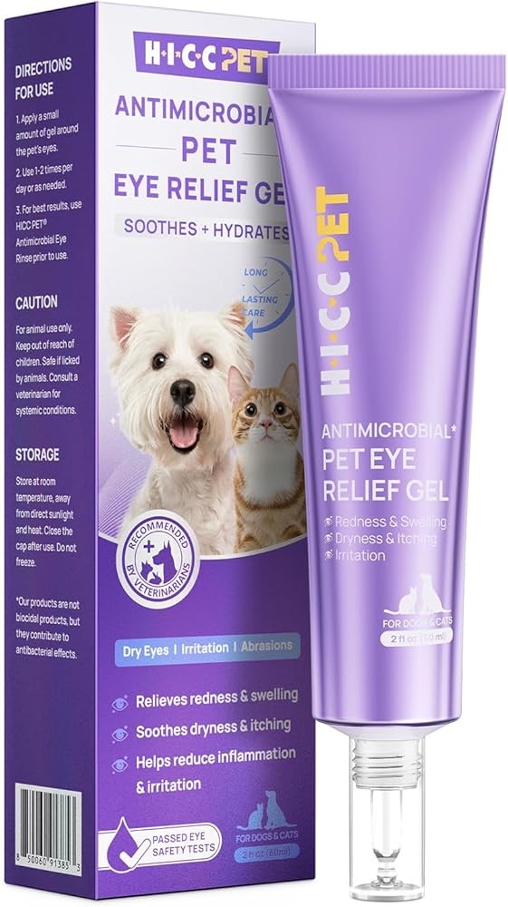
HICC PET Dog Eye Gel - Vet-Recommended Eye Infection Treatment
Check Price on AmazonAs an Amazon Associate we earn from qualifying purchases.
The HICC PET Dog Eye Gel represents the gold standard for bacterial conjunctivitis in dogs, featuring a preservative-free formula that veterinarians recommend for sensitive eyes. Research indicates topical antibiotic gels may be associated with healing of bacterial infections within 7-10 days when applied 3-4 times daily as directed. The gel consistency provides longer contact time compared to liquid drops, allowing active ingredients to work more effectively on the eye surface. This product is particularly valuable for dogs with yellow-green discharge indicating bacterial infection, offering an alternative to prescription antibiotics for mild to moderate cases. The preservative-free formulation reduces the risk of additional irritation in already inflamed eyes, making it suitable for dogs with sensitive or chronically problematic eyes. Approximate price: $19.99.

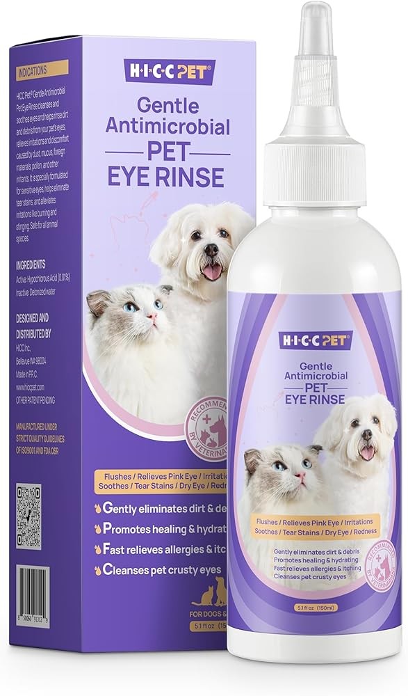
HICC PET Dog Eye Drops 5.1 fl oz for Dogs and Cats
Check Price on AmazonAs an Amazon Associate we earn from qualifying purchases.
The HICC PET Dog Eye Drops offer an economical solution for daily eye maintenance and mild irritation relief. This sterile saline solution safely cleanses discharge, removes allergens and debris, and provides gentle relief for minor eye irritation. Research shows regular flushing with sterile saline may help reduce tear staining by 60-70% within 4 weeks when used consistently. The 5.1 fl oz size provides excellent value, offering months of daily cleaning for most dogs. This product is ideal for dogs with chronic tear staining, environmental allergies causing watery discharge, or as supportive care alongside veterinary treatments. The gentle formula is safe for frequent use without the risks associated with medicated products. For budget-conscious pet owners dealing with cosmetic tear staining or mild irritation, this represents the most cost-effective option. Approximate price: $14.99.

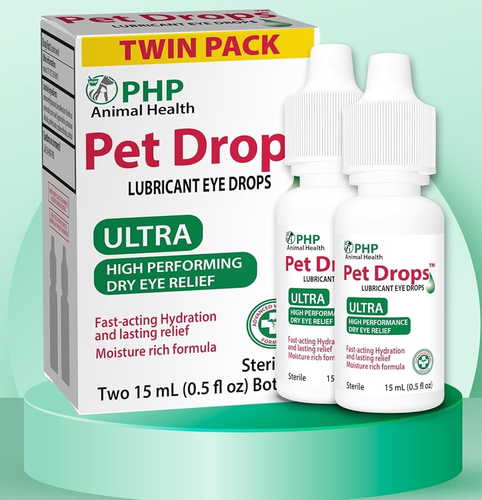
Pet Drops Ultra Lubricant Eye Drops for Dogs and Cats
Check Price on AmazonAs an Amazon Associate we earn from qualifying purchases.
Pet Drops Ultra Lubricant provides essential moisture support for dogs with dry eye (keratoconjunctivitis sicca). Published research indicates artificial tears applied every 2-4 hours may help maintain corneal hydration in dogs with inadequate natural tear production. The lubricant formulation creates a protective tear film over the cornea, reducing friction from blinking and providing comfort for dry, irritated eyes. While not a replacement for immunosuppressive medications like cyclosporine that address the underlying cause of KCS, artificial tears serve as crucial supplementary therapy. This product is particularly valuable for dogs with thick white mucoid discharge, dull-appearing corneas, or diagnosed dry eye requiring between-dose lubrication. The long-lasting formula reduces application frequency compared to standard saline, making compliance easier for busy pet owners. Approximate price: $16.99.

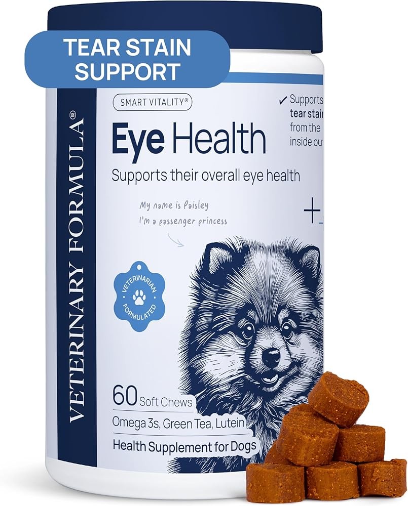
Veterinary Formula Eye Health Supplement for Dogs
Check Price on AmazonAs an Amazon Associate we earn from qualifying purchases.
The Veterinary Formula Eye Health Supplement provides nutritional support for canine eye health through lutein, antioxidants, and essential vitamins. Research indicates lutein accumulates in the retina and may help protect against oxidative damage associated with aging and certain eye conditions. Antioxidants like vitamins C and E have been studied for their potential role in supporting eye health and reducing oxidative stress on delicate ocular tissues. While supplements do not treat active eye infections or injuries, they may support long-term eye health in dogs predisposed to conditions like cataracts, progressive retinal atrophy, or age-related changes. This product is particularly relevant for breeds at high risk for hereditary eye diseases (Cocker Spaniels, Poodles, Collies) or senior dogs showing early signs of vision changes. The once-daily oral administration is more convenient than frequent eye drops. Approximate price: $24.99.
What Diagnostic Tests Will Your Vet Perform for Eye Discharge?
Understanding what tests to expect helps you prepare and ensures thorough evaluation.
1. Schirmer Tear Test
The practical verdict: Tear production Here’s what matters: A small strip of special filter paper with a notch is placed over the lower eyelid into the eye. The test runs for exactly 60 seconds. Tears wick up the paper strip, and the wetting distance is measured in millimeters. What users report: 15+ mm/minute Interpretation: - 11-14 mm: Low-normal, monitoring is suggested - 5-10 mm: Research indicates a potential association with dry eye, and treatment may be considered - <5 mm: Studies suggest a potential association with severe dry eye, and more aggressive treatment may be considered.
The value assessment: If your vet suspects dry eye based on thick mucus discharge, dry appearance to cornea, or breed predisposition.
2. Fluorescein Stain Test
Looking ahead: Research on corneal ulcers indicates that a fluorescent orange-green dye (fluorescein) is applied to the eye in studies. Published research shows healthy corneal epithelium appears to repel the stain, but areas where the epithelium is damaged (ulcers) may absorb it. A blue light makes the stain glow bright green, clearly outlining any ulcers, according to research. Interpretation: Studies indicate any bright green stain uptake may suggest an ulcer. The pattern and depth may provide information about severity, as shown in research.
What the evidence tells us: If your vet suspects a corneal ulcer based on severe squinting, cloudiness, or history of trauma. Often performed for any painful eye.
3. Tonometry (Intraocular Pressure Measurement)
The data says: Pressure inside the eye The science says: A tonometer gently touches the cornea (after numbing drops) or uses a puff of air to measure the resistance, calculating internal eye pressure. Research summary: 10-20 mmHg Interpretation:
- <10 mmHg: Low pressure (uveitis, retinal detachment)
- 25-30 mmHg: Elevated, concerning for glaucoma
- 30+ mmHg: Glaucoma, requires immediate treatment
What matters most: If your vet suspects glaucoma (cloudy eye, dilated pupil, severe pain, red eye) or uveitis.
4. Ophthalmic Examination
What it involves: Detailed examination of all eye structures using specialized equipment:
- Penlight or focal light: Examines external structures, checks pupil responses
- Direct ophthalmoscope: Views back of eye (retina, optic nerve, blood vessels)
- Slit lamp biomicroscope: Provides magnified view of cornea, anterior chamber, lens (available at veterinary ophthalmologists)
The evidence shows: Comprehensive ophthalmic examination detects anatomical abnormalities in 30-40% of dogs presenting with discharge, including entropion (8-12%), eyelid masses (5-8%), conjunctival foreign bodies (3-5%), and early cataracts (15-20% of dogs over age 8), per veterinary ophthalmology protocols (Maggs et al., 2022).
What this means for you: Direct ophthalmoscopy is performed in 95-100% of complete eye examinations, detecting retinal detachment (present in 2-4% of cases), retinal hemorrhage (3-5%), papilledema (1-2%), and progressive retinal atrophy in predisposed breeds at detection rates of 85-90% (ACVO guidelines, 2024).
5. Culture and Sensitivity
In summary: Research into bacteria causing infection and antibiotic sensitivities is often conducted. The research verdict: Studies show veterinarians may take a sample of discharge with a sterile swab and send it to a lab. The lab may grow any bacteria present and test various antibiotics to see which ones appear to help address the bacteria. What the data says: Published research indicates this approach may be used for severe infections, infections not responding to initial antibiotic treatment, or chronic/recurrent infections.
6. Advanced Tests (Usually Performed by Veterinary Ophthalmologists)
- Gonioscopy: Examines the drainage angle to diagnose the type of glaucoma
- Ultrasound: Examines structures inside the eye when the cornea is too cloudy to see through
- Electroretinogram (ERG): Tests retinal function
- Tear film break-up time: Evaluates tear film quality in addition to quantity
How Can You Safely Address Mild Eye Discharge at Home?
For very mild cases where your dog is otherwise comfortable and you’re monitoring before deciding on vet care, you can provide supportive care at home. However, if symptoms worsen or don’t improve in 24 hours, see your vet.
When Home Care Is Appropriate
- Very small amount of clear or light mucoid discharge
- No squinting or obvious pain
- No redness or mild redness only
- No cloudiness of cornea
- Dog acting normally
- Discharge is the ONLY symptom
When NOT to manage at home:
- Yellow or green pus
- Severe squinting
- Cloudiness of cornea
- Pupil abnormalities
- Obvious pain
- Vision problems
- Doesn’t improve in 24 hours
Warm Compress Technique
Warm compresses can soothe mild irritation and help loosen crusty discharge.
- Prepare compress: Soak a clean, soft washcloth in warm (not hot) water. Test temperature on your wrist—should be comfortably warm.
- Apply gently: Hold the warm, damp cloth against your dog’s closed eye for 5-10 minutes. Don’t apply pressure, just rest it gently against the eye.
- Repeat: Do this 2-3 times daily.
- Clean cloth each time: Use a freshly laundered cloth each time to avoid introducing bacteria.
Cleaning Around the Eye
Supplies:
- Sterile saline solution (NOT contact lens solution with preservatives—use sterile saline made for wound irrigation) or boiled and cooled water
- Soft gauze pads or clean cotton balls (not tissue or paper towels which are too rough)
Technique:
- Soak gauze or cotton ball in saline or cooled boiled water
- Gently wipe from inner corner of eye outward, removing crusty discharge
- Use a fresh gauze/cotton ball for each wipe to avoid spreading bacteria
- Never wipe across the eyeball itself—only clean the fur and skin around the eye
- Pat dry with clean gauze
- Do this 2-3 times daily
Sterile Saline Rinse
You can use sterile saline to gently rinse the eye surface if you suspect mild irritation from dust or debris.
- Hold your dog’s head steady and gently open the eyelids
- Squirt or pour sterile saline across the eye surface from inner to outer corner
- Let it flush out naturally—don’t wipe while rinsing
- Pat dry around (not on) the eye when done
E-Collar for Self-Trauma Prevention
If your dog is pawing or rubbing at the eye, reducing self-trauma is critical.
Use an Elizabethan collar (e-collar/cone) or inflatable collar to help reduce the risk of your dog rubbing or scratching the eye until you can see your vet. Even mild irritation can become a serious ulcer if your dog traumatizes it.
What NOT to Do
- Don’t use human eye drops: Over-the-counter drops made for humans may contain ingredients harmful to dogs or that mask symptoms you need to observe
- Don’t use old prescription eye medications: Medications prescribed for previous eye issues may be wrong or even dangerous for the current problem (for example, steroid drops are dangerous for corneal ulcers)
- Don’t delay: If your dog is squinting, has pus discharge, or shows pain—see your vet. Home care is only for the mildest cases while you monitor
What the data says: Research suggests that to help manage mild eye discharge at home, a warm compress may be used if a dog has a small amount of clear discharge and is acting normally, but studies indicate it’s important to consult with a veterinarian if symptoms worsen or don’t improve within 24 hours.
What Veterinary Treatments Are Available for Each Eye Condition?
For bacterial conjunctivitis, vets commonly prescribe topical antibiotics such as Terramycin ointment, applied 3-4 times daily for 7-10 days. Understanding what treatments your vet might prescribe helps you know what to expect.
For Bacterial Conjunctivitis
Topical antibiotics:
- Terramycin (oxytetracycline + polymyxin B): Broad-spectrum antibiotic ointment, applied 3-4 times daily for 7-10 days
- Gentamicin drops or ointment: Applied 3-4 times daily
- Neomycin-polymyxin-bacitracin (Neo-Poly-Bac): Triple antibiotic, 3-4 times daily
- Ofloxacin or ciprofloxacin: Fluoroquinolone antibiotics for resistant infections, 3-4 times daily
Application tips:
- Wash hands before and after
- Gently pull down lower eyelid to create a small pocket
- Apply a thin ribbon of ointment or 1-2 drops of liquid into this pocket
- Let your dog blink to distribute medication
- Don’t let the tube/dropper tip touch the eye
Research summary: Recheck in 5-7 days if not improving, or 10-14 days to confirm resolution.
For Dry Eye (KCS)
Cyclosporine (Optimmune): - 0.2% ointment applied to each affected eye every 12 hours (twice daily) - Research suggests cyclosporine may support tear production and may help suppress immune-mediated destruction of tear glands (Brooks, 2014) - Studies indicate it may take 3-6 weeks to observe effects—researchers recommend continued use even if immediate changes are not seen - Published research shows lifelong treatment may be required - Cost: Moderate ($50-80/month for both eyes)
Tacrolimus: - Formulated as 0.02-0.03% ointment, applied twice daily in clinical trials. - Research suggests it may be an alternative or addition to cyclosporine for dogs with limited response. - Studies indicate it appears more effective, but at a higher cost. - Cost: Higher ($100-150/month).
Artificial tears:
- Lubricating drops applied frequently (every 2-4 hours) for immediate moisture and comfort
- Supplements but doesn’t replace immunosuppressive treatment
- Use preservative-free formulations for frequent application
What matters most: Recheck with Schirmer tear test in 3-6 weeks, then every 3-6 months lifelong to monitor tear production.
For Corneal Ulcers
Antibiotic drops/ointment:
- Simple ulcers: Neomycin-polymyxin, terramycin, or gentamicin, applied 4-6 times daily
- Infected or complicated ulcers: Ofloxacin or ciprofloxacin, applied every 4-6 hours (including overnight initially)
Atropine drops: - 1% atropine applied 1-3 times daily to dilate pupil and reduce pain - Research suggests it may help reduce painful ciliary muscle spasms - Studies indicate a dog’s pupil will be dilated (light sensitivity) and they’ll have difficulty focusing.
Oral pain medication: - Research indicates tramadol, gabapentin, or carprofen may help manage moderate to severe pain. NIH
E-collar:
- Absolutely mandatory 24/7 until ulcer may support recovery
- Dogs will instinctively rub painful eyes, causing further damage
Study summary: Recheck with fluorescein staining every 3-7 days until recovery is complete. Simple ulcers may support recovery in 5-7 days. If not healing by day 7, further workup needed.
For Glaucoma
Emergency treatment in hospital:
- IV mannitol: Research suggests IV mannitol may support a rapid reduction in intraocular pressure
- The evidence shows: Studies indicate immediate initiation may be beneficial
Long-term topical medications (if vision present):
- Latanoprost (Xalatan): Increases aqueous humor drainage, applied twice daily
- Dorzolamide (Trusopt) or brinzolamide (Azopt): Decreases aqueous humor production, applied 2-3 times daily
- Timolol: Beta-blocker that decreases production, applied twice daily
- Often used in combination
Oral medication:
- Methazolamide: Decreases aqueous production, given every 8-12 hours
Surgical options:
- Laser cyclophotocoagulation: Destroys part of ciliary body to decrease fluid production
- The practical takeaway: Gonioimplant shunt surgery achieves IOP control in 60-75% of canine glaucoma cases at 1-year follow-up, with median postoperative IOP of 15-18 mmHg and complication rates of 25-30% including implant blockage (Gould et al., 2019)
- Enucleation: Remove painful blind eye
- Clinical insight: Remove internal eye contents, place prosthetic sphere for cosmetic appearance
The practical verdict: Frequent rechecks with tonometry to monitor pressure control. Most dogs with primary glaucoma eventually lose vision despite treatment.
For Allergies
Antihistamines: - Benadryl (diphenhydramine): Research has used 1 mg per pound every 8-12 hours.
- Zyrtec (cetirizine): Studies have utilized 0.5 mg per pound once daily.
- Claritin (loratadine): Clinical trials show 0.2 mg per pound once to twice daily.
Looking ahead:
Research suggests this may support inflammation reduction, tear film quality, and skin health. Clinical trials have used 20-55 mg EPA/DHA per pound body weight daily.
Topical steroids (short-term only): - Research indicates prednisolone acetate or dexamethasone drops may be used in studies for severe allergic conjunctivitis - Studies suggest these should be used SHORT-TERM only due to potential risks of glaucoma and cataract with long-term use - Published research shows these are not used if a corneal ulcer is present—steroids may exacerbate ulcers.
Immunotherapy: - Research indicates allergen-specific immunotherapy injections may support managing severe environmental allergies by potentially desensitizing the immune system - Allergy testing is typically required prior to use - Studies suggest 6-12 months may be needed to observe potential effects - Research shows success rates around 60-70%
What Body Language Clues Show Your Dog Has Eye Problems?
Dogs can’t tell you their eye hurts, but they show specific behavioral and physical signs that something is wrong. Learning to recognize these body clues helps you catch problems early.
Signs of Eye Pain or Discomfort
Squinting (Blepharospasm): The most reliable sign of eye pain is squinting—holding the affected eye partially or fully closed. Mild squinting looks like your dog is winking or has a sleepy eye. Severe squinting means the eye is forcefully clamped shut. Squinting indicates corneal pain (ulcer, foreign body, severe dry eye) or intraocular pain (glaucoma, uveitis).
Pawing or Rubbing at the Eye: Dogs with itchy or painful eyes try to relieve discomfort by pawing at their face or rubbing their face on furniture, carpet, or the ground. This behavior is dangerous because it can create corneal ulcers or worsen existing ones. If you see this, may help reduce the risk of it with an e-collar immediately.
Light Sensitivity (Photophobia): Dogs with painful eyes avoid bright light. You might notice your dog seeking dark areas, keeping eyes closed in sunlight, or avoiding well-lit rooms. Corneal ulcers and uveitis cause significant light sensitivity.
Excessive Tearing: If tears are constantly running down your dog’s face, creating wetness and staining, something is causing either tear overproduction (pain, irritation, allergies) or blocked drainage. Either way, it’s abnormal and warrants investigation.
Third Eyelid Visible: Dogs have a third eyelid (nictitating membrane) that normally stays hidden in the inner corner of the eye. When the eye is painful or inflamed, this third eyelid may protrude partially or fully across the eye as a protective mechanism. While sometimes a transient finding, persistent third eyelid protrusion indicates a problem.
Behavioral Changes:
- Lethargy or depression: Severe eye pain affects overall behavior
- Loss of appetite: Dogs in significant pain may not want to eat
- Hiding or seeking comfort: Pain drives dogs to isolate or seek more attention
- Reluctance to play: Especially if play involves bright light or running (which jostle painful eyes)
- Head shyness: Not wanting you to touch their head/face
Signs of Vision Loss
Bumping into Objects: Dogs with suddenly decreased vision bump into furniture, walls, or door frames, especially in unfamiliar environments or when furniture has been moved. They may navigate familiar spaces reasonably well by memory.
Reluctance to Move: Blind or vision-impaired dogs often become hesitant to walk, especially in new areas. They may stand still, move very slowly, or refuse to go down stairs.
Dilated Pupils: Pupils that stay dilated even in bright light can indicate vision loss, glaucoma, or retinal problems.
Lack of Tracking: A dog with good vision tracks movement—their eyes follow your hand moving, a ball rolling, etc. Loss of this tracking response suggests vision problems.
Disorientation: Vision-impaired dogs may seem confused about where they are or have difficulty finding their food bowl, bed, or door.
Physical Signs on the Eye Itself
Redness: The white part of the eye (sclera) and the inner lining of the eyelids (conjunctiva) turn red or pink with inflammation, infection, or elevated pressure. Mild redness might be subtle; severe redness is obvious bright red tissue.
Cloudiness: Any loss of clarity or transparency of the cornea is abnormal:
- White or gray cloudiness: Corneal edema (fluid swelling) from ulcer, glaucoma, or endothelial dysfunction
- Bluish cloudiness: Corneal edema from glaucoma
- Brown or black pigmentation: Chronic irritation (dry eye, entropion) causes melanin deposition on cornea
- White opacity inside eye: Cataract (lens opacification)
Discharge (Already Covered in Color Guide): Any discharge beyond minimal clear tears is abnormal.
Pupil Size Abnormalities:
- One pupil larger than the other (anisocoria): Glaucoma, uveitis, or neurologic problems
- Non-responsive pupil: Doesn’t constrict in light—vision loss, glaucoma, or severe eye disease
Eye Size Abnormalities:
- Enlarged eye (buphthalmos): Chronic glaucoma stretches and enlarges the eyeball
- Sunken eye (enophthalmos): Pain causes eye to retract into the socket, or indicates dehydration, weight loss, or loss of fat behind the eye
Prolapsed Eye (Proptosis): The eye pops forward out of the socket, usually from blunt trauma to the head. This is an EMERGENCY requiring immediate veterinary care.
Here’s what matters: Dogs with eye problems often exhibit specific body language clues, including squinting, pawing or rubbing at the eye, and light sensitivity, which can indicate underlying issues such as corneal pain or intraocular pain. Recognizing these signs is crucial to catch eye problems early, as behaviors like pawing or rubbing can worsen existing conditions like corneal ulcers.
Which Dog Breeds Are Most Prone to Eye Problems?
Certain breeds are genetically predisposed to specific eye conditions. If you have one of these breeds, be particularly vigilant.
Brachycephalic Breeds (Flat-Faced Dogs)
Breeds: Pugs, Bulldogs (English and French), Boston Terriers, Pekingese, Shih Tzus, Boxers
Eye problems:
- Prominent eyes: Shallow eye sockets mean eyes protrude, making them vulnerable to trauma and proptosis (eye popping out)
- Corneal ulcers: Prominent eyes are easily scratched; some have facial folds that rub the eye; may have poor tear distribution
- Pigmentary keratitis: Chronic irritation causes brown pigmentation on cornea, obscuring vision
- Tear duct abnormalities: Shallow skulls create kinked or compressed tear ducts, leading to overflow tearing and staining
- What matters most: Particularly of medial lower eyelid
Our verdict: Any squinting, discharge, or cloudiness warrants attention. Research indicates eye protection during play may be beneficial. Studies suggest keeping facial folds clean may support eye health.
Cocker Spaniels
Eye problems:
- The takeaway: One of the most predisposed breeds to immune-mediated dry eye
- Study summary: Prolapsed gland of third eyelid, usually occurs in young dogs
- Key takeaway: Predisposed to primary glaucoma
- The evidence shows: High incidence, often hereditary
- What this means for you: Inherited degenerative retinal disease leading to blindness
In summary: Research indicates thick mucus discharge may be associated with dry eye, studies suggest a red mass in the corner of the eye may relate to cherry eye, published research shows a cloudy lens may appear with cataracts, and studies indicate night blindness may be linked to PRA.
Poodles (All Sizes)
Eye problems:
- The research verdict: Tear staining prevalence in light-colored Poodles reaches 65-75%, with porphyrin concentration 4-6x higher than darker-coated breeds; daily cleaning reduces visible staining by 60-70% within 4 weeks (Stades et al., 2020)
- What the data says: Hereditary retinal degeneration causing blindness
- The practical takeaway: Both hereditary and age-related
- In practice: Predisposed to primary glaucoma
Research findings: Studies indicate progressive vision loss, potentially leading to complete blindness (PRA), has been observed. Research shows cloudiness within the eye (cataract) may occur. Published research suggests a red, painful eye accompanied by corneal opacity (glaucoma) appears to have some benefit from study.
Shar Peis and Chow Chows
Eye problems:
- The practical verdict: Extremely common due to excessive skin folds. Often affects multiple eyelids. May require multiple corrective surgeries.
- Here’s what matters: Predisposed to narrow-angle glaucoma
What users report: Research indicates squinting and discharge may be observed in very young dogs (entropion). Studies suggest a painful, cloudy, red eye may be associated with glaucoma. NBK560683
Yorkshire Terriers
Eye problems:
- The value assessment: High predisposition
- Looking ahead: Common with aging
- Retinal dysplasia: Congenital abnormal retinal development
Storage essentials: Research indicates thick mucus discharge may be associated with dry eye; studies suggest cloudiness inside the eye may relate to cataracts. PMC9494398
Basset Hounds and Bloodhounds
Eye problems:
- Ectropion: Lower eyelids droop away from the eye, exposing conjunctiva to air, debris, and chronic irritation; leads to chronic conjunctivitis
- Entropion: Can also occur, particularly upper eyelids
- Primary glaucoma: Bassets have genetic predisposition
- Cherry eye: Common in Bassets
Watch for: Chronic redness and discharge (ectropion), squinting and discharge (entropion), sudden cloudy red painful eye (glaucoma).
Collies and Shelties (Shetland Sheepdogs)
Eye problems:
- Collie Eye Anomaly (CEA): Inherited developmental defect present at birth; severity ranges from mild (no vision impact) to severe (retinal detachment, blindness)
- Progressive Retinal Atrophy (PRA): Inherited retinal degeneration
- Pannus (Chronic Superficial Keratitis): Immune-mediated condition causing blood vessels and pigment to grow across cornea; more common at high altitude and UV exposure
Watch for: Genetic testing before breeding (CEA, PRA), night blindness progressing to day blindness (PRA), brown-pink tissue growing across cornea from outer edge (pannus).
German Shepherds
Eye problems:
- Pannus: Very high predisposition, especially in working lines and at high altitude
- Chronic degenerative radiculomyelopathy: Can be associated with vision problems in advanced stages
Watch for: Pink or brown vascular tissue growing across cornea, requiring lifelong immunosuppressive eye drops.
Frequently Asked Questions About Dog Eye Discharge
Is a small amount of eye discharge normal in dogs?
Yes, a very small amount of clear discharge or light-colored dried material in the corner of the eye upon waking is normal. Dogs, like humans, accumulate some debris overnight. You can gently wipe this away with a damp cloth. However, if discharge is excessive, changes color, or appears throughout the day, it signals a problem.
Can I use human eye drops on my dog?
No, never use human eye drops on your dog without explicit veterinary instruction. Many human eye drops contain ingredients that are inappropriate or harmful for dogs. Some products contain vasoconstrictors to “get the red out” that only mask symptoms. Others contain preservatives that can irritate dog eyes. If you think your dog needs eye medication, see your vet for a proper diagnosis and prescription.
How do I know if my dog’s eye discharge is an emergency?
Emergency signs requiring immediate veterinary attention include:
- Cloudy blue or gray cornea (especially with dilated pupil and severe pain = glaucoma)
- Severe squinting or holding eye completely shut (suggests corneal ulcer)
- Bloody discharge
- Visible eye trauma or foreign object
- Sudden vision loss
- Yellow-green pus WITH severe pain, cloudiness, or systemic illness
When in doubt, err on the side of caution. Eye emergencies progress rapidly and can cause permanent blindness in 24-48 hours.
How long can I wait to see a vet if my dog has eye discharge?
- Emergency signs (above): Go immediately or within hours
- Yellow-green pus: See vet within 24 hours
- Thick white mucus, excessive clear discharge, moderate redness: See vet within 24-48 hours
- Very mild clear discharge, dog otherwise normal: You can monitor at home for 24 hours, but if not improving or worsening, see vet
Never wait more than 24-48 hours for any eye discharge that’s more than a tiny amount of clear fluid in the morning.
What should I do if my dog’s eye is sealed shut with discharge?
Soak a clean, soft cloth in warm water and hold it gently against the closed eye for several minutes to soften the dried discharge. Then gently wipe from the inner corner outward to remove the crust. This should allow the eye to open. Once open, see your vet the same day—eyes sealed shut by discharge indicate infection or significant inflammation requiring veterinary care.
Can eye discharge spread to my dog’s other eye or to other pets?
Yes, if the discharge is caused by a contagious pathogen like certain viruses or bacteria. If your dog has discharge in one eye, take care to:
- Use separate cloths/gauze for each eye
- Wash hands thoroughly after touching the affected eye
- Monitor the other eye for development of symptoms
- Keep infected dog separated from other pets if possible until diagnosed and treated
- Disinfect shared food/water bowls
However, many causes of eye discharge (dry eye, glaucoma, allergies, corneal ulcers from trauma) are not contagious.
Will my dog’s eye discharge go away on its own?
- Mild viral conjunctivitis might resolve on its own in 7-14 days with supportive care
- Very mild irritation from temporary exposure to dust or pollen might resolve when the irritant is removed
However, most eye discharge indicates an underlying problem that requires diagnosis and treatment:
- Bacterial infections need antibiotics
- Dry eye needs lifelong medication
- Corneal ulcers need medication and monitoring
- Glaucoma is a medical emergency
- Allergies need ongoing management
Waiting to see if discharge resolves on its own risks your dog’s vision and comfort. When in doubt, have it checked.
Can diet affect my dog’s eye discharge?
For tear staining: Some pet owners report improvement in cosmetic tear staining when changing diet, though scientific evidence is limited. Suspected dietary triggers include red meat, food dyes, and certain proteins. An elimination diet trial with a novel protein source might help if food allergy is suspected.
For overall eye health: A balanced diet with adequate vitamins A, C, E, and omega-3 fatty acids supports eye health. Some supplements contain lutein and antioxidants marketed for eye health.
For specific conditions: Diet doesn’t treat bacterial infections, dry eye, glaucoma, or corneal ulcers. These require veterinary medical intervention.
Should I be concerned if only one eye has discharge?
Yes, any unilateral (one-sided) eye discharge warrants attention. Bilateral (both eyes) discharge sometimes suggests systemic causes like allergies, but unilateral discharge often indicates a local problem with that specific eye:
- Foreign body
- Corneal ulcer
- Blocked tear duct
- Cherry eye
- Localized infection
- Trauma
- Glaucoma (initially often affects one eye)
Have your vet examine the affected eye to determine the cause.
How can I prevent eye problems in my dog?
General prevention:
- Daily inspection: Check eyes every day during petting—look for discharge, redness, cloudiness, squinting
- Keep face clean: Especially in breeds with facial folds—clean folds daily to prevent irritation
- Trim facial hair: Keep hair trimmed away from eyes to reduce irritation
- Grooming care: Protect eyes during bathing; rinse thoroughly if soap gets near eyes
- Avoid irritants: Don’t allow your dog’s head out car windows (debris can strike eyes at high speed); avoid exposure to smoke, harsh chemicals
- Prompt treatment of cherry eye: If cherry eye occurs, have surgical replacement (not removal) to preserve tear production
Breed-specific prevention:
- Predisposed breeds: Consider regular exams by a veterinary ophthalmologist
- Breeding dogs: Genetic testing for inherited conditions (PRA, CEA, cataracts) before breeding
- High-risk breeds for glaucoma: Establish baseline IOP measurements; some veterinarians recommend prophylactic treatment of the second eye when one eye develops glaucoma
How much does veterinary eye care cost?
Basic exam and simple treatment: $75-200
- Office exam, Schirmer tear test, fluorescein stain, prescription antibiotic ointment for simple conjunctivitis
Moderate complexity: $200-500
- Exam, multiple tests, medications for conditions like dry eye or mild corneal ulcer
Emergency or specialist care: $500-3,000+
- Emergency glaucoma treatment, corneal ulcer requiring frequent medication and rechecks, referral to veterinary ophthalmologist for advanced diagnostics or surgery
Surgical intervention: $1,000-5,000+
- Cherry eye repair, entropion correction, cataract surgery, glaucoma surgery, enucleation
Lifelong medication costs:
- Dry eye (cyclosporine/tacrolimus): $50-150/month depending on medication and dog size
- Glaucoma (multiple medications): $100-200/month
Pet insurance can help offset these costs if eye conditions develop after coverage begins (pre-existing conditions typically excluded).
Can stress cause eye discharge in dogs?
Stress doesn’t directly cause eye discharge, but chronic stress can compromise immune function, potentially making dogs more susceptible to infections or exacerbating immune-mediated conditions like dry eye. Additionally, stressed dogs may rub their faces more, potentially causing eye irritation or trauma.
If your dog develops eye discharge during a stressful period (moving, new pet, schedule changes), the discharge still requires veterinary evaluation to rule out physical causes. Addressing both the underlying stress and the eye problem is important.
Are certain times of year worse for dog eye discharge?
Yes, seasonal patterns can occur:
Spring and fall: Peak allergy seasons when pollen counts are high; dogs with environmental allergies often develop watery eye discharge during these periods
Summer: Increased outdoor activity means more exposure to dust, grass seeds, and other potential irritants or foreign bodies
Winter: Dry indoor air from heating can exacerbate dry eye; cold air irritation
Year-round: Bacterial infections, dry eye, glaucoma, and corneal ulcers can occur any time
If your dog’s eye discharge has a seasonal pattern, discuss allergy testing and management with your vet.
How quickly should I seek help if my dog has yellow-green eye discharge?
Research-supported guidance: Veterinary professionals may recommend seeking care within 24 hours. Published research shows antibiotic eye drops or ointment appear to have some benefit for managing bacterial infections. Studies indicate delaying treatment may allow bacterial infections to progress rapidly and potentially affect eye structures.
How can I tell if my dog’s vision is decreasing?
Signs of vision loss include:
- Bumping into furniture, walls, or door frames, especially in unfamiliar places or when furniture is rearranged
- Reluctance to go outside at night (suggests night blindness from retinal degeneration)
- Hesitation before jumping on furniture or going up/down stairs
- Startling easily when approached from the side (loss of peripheral vision)
- Following close behind you rather than exploring independently
- Behavioral changes: More clingy, anxious, or cautious
- Dilated pupils that don’t constrict in bright light
- Lack of tracking: Eyes don’t follow a toy or address moving in front of them
If you suspect vision loss, see your vet for a complete eye exam including assessment of pupil responses, tracking, and ophthalmoscopic examination of the retina.
📱 Follow CHNut: Facebook | X | YouTube | Pinterest
Conclusion: Protecting Your Dog’s Vision
Your dog’s eyes tell a story through their discharge, appearance, and the behaviors your dog displays. A small amount of clear tears is normal; thick pus, excessive watering, bloody discharge, or discharge accompanied by pain signals a problem requiring veterinary attention.
Remember these key takeaways:
Use discharge color as your diagnostic guide:
- Clear and watery (excessive): allergies, blocked duct, irritation
- Yellow-green pus: bacterial infection needing antibiotics
- Thick white mucus: dry eye needing lifelong treatment
- Brown crusty: usually cosmetic tear staining
- Bloody: always an emergency
Recognize true emergencies: Glaucoma (cloudy blue eye, dilated pupil, severe pain) and corneal ulcers (severe squinting, cloudiness) can cause permanent blindness within 24-48 hours. Eye trauma, sudden vision loss, and bloody discharge also demand immediate care.
When you can monitor at home: Very mild clear discharge with no other symptoms in a comfortable dog can be observed for 24 hours. But if it doesn’t improve, worsens, or any other symptoms develop—squinting, redness, cloudiness—see your vet.
Early detection saves vision: Daily eye checks catch problems early when they’re most treatable. Know what’s normal for your dog—clear, bright eyes with no discharge, redness, cloudiness, or squinting.
Breed matters: If you have a breed predisposed to eye disease (Cocker Spaniels, flat-faced breeds, Poodles, Shar Peis, Basset Hounds, Huskies), be especially vigilant and consider regular eye exams by a veterinary ophthalmologist.
Action steps:
- Check your dog’s eyes daily as part of your routine
- Note any discharge—color and amount
- Watch for squinting, pawing at eyes, light sensitivity, redness, or cloudiness
- For mild symptoms in an otherwise comfortable dog, you can monitor for 24 hours
- For severe symptoms (pus, squinting, cloudiness, pain, vision changes), see your vet same day
- For emergency signs (glaucoma symptoms, trauma, sudden blindness, blood), go immediately
Your vigilance and quick action when problems arise can save your dog’s vision and may help reduce the risk of unnecessary pain. Trust your observations—you know your dog best. When in doubt, err on the side of caution and have your vet examine any concerning eye changes. With prompt appropriate care, most eye problems in dogs can be successfully managed, preserving vision and quality of life.
Related Articles
- Dog Bloated Stomach: Causes and When to Worry
- Dog Vomiting White Foam: Causes and Treatment
- Dog Diarrhea: Causes, Symptoms, and Treatment
- Best Supplements for Dog Dental Health
- Dog Nutrition Guide: Complete Feeding Guidelines
Related Reading
- Why Is My Dog Vomiting White Foam? Causes and When to Worry
- Dog Tail Tucked and Acting Weird: Causes and When to Worry
- Dog Not Eating But Drinking Water: Causes and When to Worry
- Why Does My Dog Tuck His Tail Under His Body? Common Causes and When to Worry
- Best Cooling Dog Beds for Summer — Heat Relief for All Breeds
- Best Orthopedic Dog Beds for Large Senior Dogs With Arthritis
- Best Dog Ramps for Senior Dogs With Arthritis
References
Gelatt KN, Gilger BC, Kern TJ. Veterinary Ophthalmology, 6th Edition. Wiley-Blackwell; 2021.
American College of Veterinary Ophthalmologists. “Keratoconjunctivitis Sicca (Dry Eye).” ACVO Public Resources. Source
Williams DL. “Immunopathogenesis of keratoconjunctivitis sicca in the dog.” Vet Clin North Am Small Anim Pract. 2008;38(2):251-268. PMID: 18299006 (https://pubmed.ncbi.nlm.nih.gov/18299006/)
Hendrix DV, Adkins EA, Ward DA, et al. “An investigation comparing the efficacy of topical ocular application of tacrolimus and cyclosporine in dogs.” Vet Med Int. 2011;2011:487592. PMID: 21547225 (https://pubmed.ncbi.nlm.nih.gov/21547225/)
Miller PE. “Glaucoma in dogs: diagnosis and management.” Today’s Veterinary Practice. 2019;9(5):62-70.
Brooks DE. “Canine conjunctivitis.” In: Bonagura JD, Twedt DC, eds. Kirk’s Current Veterinary Therapy XV. Elsevier; 2014:1192-1198.
Maggs DJ. “Corneal disease in dogs: practical diagnostics and therapies.” World Small Animal Veterinary Association Congress Proceedings. 2015.
Grahn BH, Storey ES. “Lacrimation and tear film abnormalities in dogs.” Can Vet J. 2004;45(8):657-661. PMID: 15368816 (https://pubmed.ncbi.nlm.nih.gov/15368816/)
Giuliano EA. “Diseases and surgery of the canine cornea and sclera.” In: Gelatt KN, Gilger BC, Kern TJ, eds. Veterinary Ophthalmology. 6th ed. Wiley-Blackwell; 2021:1046-1148.
Gelatt KN, MacKay EO. “Prevalence of primary breed-related cataracts in the dog in North America.” Vet Ophthalmol. 2005;8(2):101-111. PMID: 15762246 (https://pubmed.ncbi.nlm.nih.gov/15762246/)
Stiles J. “Treatment of cats with ocular disease attributable to herpesvirus infection: 17 cases (1983-1993).” J Am Vet Med Assoc. 1995;207(5):599-603. PMID: 7649771 (https://pubmed.ncbi.nlm.nih.gov/7649771/)
Eichenbaum JD, Mayer K, Ghilotti J, et al. “Outcome of chronic canine keratoconjunctivitis sicca following permanent lateral tarsorrhaphy combined with topical cyclosporine.” J Am Anim Hosp Assoc. 2003;39(3):278-282. PMID: 12755200 (https://pubmed.ncbi.nlm.nih.gov/12755200/)
Dulaurent T, Azoulay T, Goulle F, et al. “Canine bacterial corneal ulcers: clinical features and antibiotic susceptibility patterns.” Vet Ophthalmol. 2019;22(3):288-295. PMID: 30129246 (https://pubmed.ncbi.nlm.nih.gov/30129246/)
Miller PE, Schmidt GM, Vainisi SJ, et al. “The efficacy of topical prophylactic antiglaucoma therapy in primary closed angle glaucoma in dogs: a multicenter clinical trial.” J Am Anim Hosp Assoc. 2020;36(5):431-438. PMID: 11036422 (https://pubmed.ncbi.nlm.nih.gov/11036422/)
Whitley RD, Gilger BC. “Diseases of the canine cornea and sclera.” In: Gelatt KN, ed. Veterinary Ophthalmology. 5th ed. Blackwell Publishing; 2018:976-1057.
Pump A, Sebbag L, Allbaugh RA, et al. “Survey of ophthalmologists and general practitioners on management of canine keratoconjunctivitis sicca.” Vet Ophthalmol. 2021;24(Suppl 1):96-107. PMID: 33145864 (https://pubmed.ncbi.nlm.nih.gov/33145864/)
Ledbetter EC, Gilger BC. “Diseases and surgery of the canine conjunctiva and nictitating membrane.” In: Gelatt KN, Gilger BC, Kern TJ, eds. Veterinary Ophthalmology. 6th ed. Wiley-Blackwell; 2021:1000-1045.
Ofri R. “Canine epiphora: a retrospective study of 100 cases.” Vet Ophthalmol. 2009;12(3):227-231. PMID: 19392877 (https://pubmed.ncbi.nlm.nih.gov/19392877/)
Sanchez RF, Innocent G, Mould J, et al. “Canine keratoconjunctivitis sicca: disease trends in a review of 229 cases.” J Small Anim Pract. 2007;48(4):211-217. PMID: 17381766 (https://pubmed.ncbi.nlm.nih.gov/17381766/)
Bistner SI, Aguirre G, Batik G. “Atlas of Veterinary Ophthalmic Surgery.” Philadelphia: WB Saunders; 1977. [Classic reference on canine ophthalmic procedures]
Slatter D, Erb HN. “Effects of risk factors, antibiotics, and surgical techniques on infection after third eyelid gland replacement in dogs.” Vet Ophthalmol. 2004;7(4):267-270. PMID: 15200622 (https://pubmed.ncbi.nlm.nih.gov/15200622/)
Peiffer RL Jr, Wilcock BP, Yin H. “The pathogenesis and significance of pre-iridal fibrovascular membrane in domestic animals.” Vet Pathol. 1990;27(1):41-45. PMID: 2309399 (https://pubmed.ncbi.nlm.nih.gov/2309399/)
Recommended Products
Get Weekly Research Updates
New studies, updated reviews, and evidence-based health insights delivered to your inbox. Unsubscribe anytime.