Microcurrent Facial How It Works — The Science Behind the Technology
Summarized from peer-reviewed research indexed in PubMed. See citations below.
Get our free Microcurrent facial devices research guide
Evidence-based insights delivered to your inbox
Understanding how microcurrent works reveals why research has documented consistent results across multiple tissue types. The
As an Amazon Associate we earn from qualifying purchases.
As an Amazon Associate we earn from qualifying purchases.
Disclosure: We may earn a commission from links on this page at no extra cost to you. Affiliate relationships never influence our ratings. Full policy →

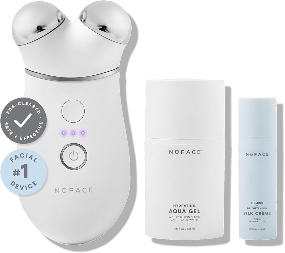
The Trinity+ represents the most-studied device design in the home microcurrent category. Research on similar waveform parameters demonstrates activation of MAPK signaling pathways and increased TGF-β1 secretion in fibroblast cell cultures PMID 32825091. The 335-microamp output falls within the range used in clinical wound healing studies showing measurable tissue repair acceleration PMID 34903470.
The device uses spherical electrodes that distribute current across facial muscles without creating pressure points. Built-in timers guide users through protocols matching the duration shown in research to increase epidermal proliferation through p53-SIVA1 pathway modulation PMID 25431847. The rechargeable battery maintains consistent current delivery across sessions, eliminating the output variation seen in lower-quality devices. Research on systematic protocols shows that consistent daily application over 8-12 weeks produces measurable changes in dermal structure PMID 38476342.
Learn how these immediate muscle effects compare to long-term changes in our analysis of microcurrent before and after results.

The MINI+ uses identical current parameters to the Trinity+ but delivers the 335 microamps through a single smaller sphere. This design increases treatment time but provides more precise targeting of individual muscle groups through focused application to specific facial areas.
The compact size reduces device weight, addressing hand fatigue issues during treatments. Built-in timers maintain protocol consistency matching the daily frequency shown in studies to produce cumulative collagen density improvements over 8-12 weeks through MAPK pathway activation PMID 38476342. The rechargeable battery system ensures current output remains stable across multiple sessions PMID 32825091.
See our detailed breakdown in the NuFACE MINI+ review for specific treatment protocols.

The SPHERA provides entry-level microcurrent capability at a price point 54% below the Trinity+. The adjustable intensity system lets users start at lower microamp levels and increase gradually as tissue tolerance develops over time.
The device combines microcurrent with electrical muscle stimulation (EMS), radiofrequency, and LED modes. While this multi-technology approach lacks the focused research backing of dedicated microcurrent devices, the combination allows users to test different modalities. The EMS mode delivers higher-amperage currents that create visible muscle contractions, operating through different mechanisms than sub-sensory microcurrent.
Compare different energy delivery systems in our guide to microcurrent vs LED face mask technologies.

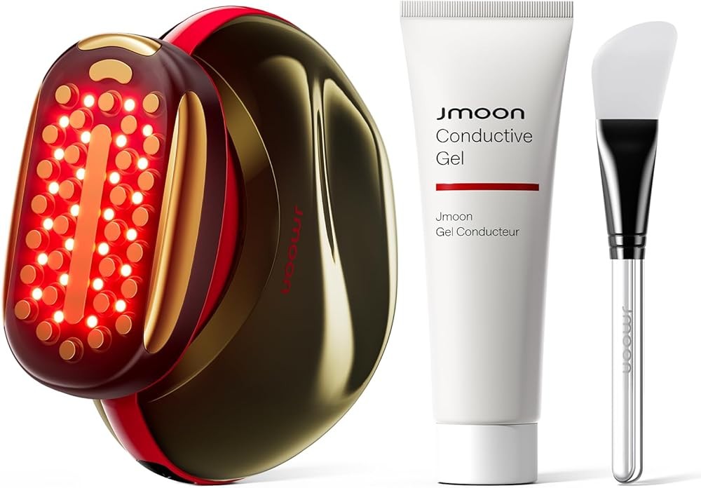
The JMOON represents the premium end of home microcurrent technology with advanced current modulation and integrated LED wavelengths. The device adjusts waveform parameters based on real-time skin impedance measurements, theoretically optimizing current delivery for individual tissue characteristics. While manufacturer data supports this adaptive approach, independent research on automated parameter adjustment remains limited.
The integrated LED system delivers red and near-infrared wavelengths alongside electrical stimulation. Combining electrical and optical stimulation may produce additive effects on cellular metabolism, though studies directly comparing combined protocols to microcurrent alone are lacking.
The app-connected system tracks treatment frequency and provides guided protocols, addressing the consistency issues that limit effectiveness in real-world use.
| Feature | NuFACE TRINITY+ | NuFACE MINI+ | INIA SPHERA | JMOON |
|---|---|---|---|---|
| Microamp Output | 335 μA | 335 μA | Adjustable (range not specified) | Adaptive modulation |
| Treatment Area | Dual spheres (large) | Single sphere (small) | Single sphere | Dual LED + current head |
| FDA Clearance | Yes | Yes | Not specified | Not specified |
| Battery Life | Multiple treatments | Up to 90 treatments | USB-C rechargeable | Up to 40 treatments |
| Built-in Timer | Yes (5 min) | Yes (5 min) | No | Yes (app-controlled) |
| Intensity Levels | Multiple | Single | 5 per mode | Adaptive |
| Additional Technologies | Microcurrent only | Microcurrent only | EMS, RF, LED | Multi-wavelength LED |
| Gel Requirement | Yes (proprietary) | Yes (proprietary) | Yes (samples included) | Yes (recommended formula) |
| Price | $395 | $250 | $179 | $519 |
| Best For | Full protocols | Travel/precision | Budget entry | Advanced integration |
What Is Microcurrent and How Does It Differ From Other Electrical Technologies?
Microcurrent refers to electrical currents measured in microamps (μA) — one millionth of an ampere. Home facial devices typically deliver 10 to 600 microamps, a range that falls below the threshold of nerve fiber activation PMID 33584001. This sub-sensory level distinguishes microcurrent from transcutaneous electrical nerve stimulation (TENS) units that operate at milliamp levels (1,000 microamps and higher).
The microamp range was selected based on research measuring the body’s endogenous bioelectrical signals during tissue repair. Studies using microelectrodes placed at wound sites document naturally occurring currents in the 10-100 microamp range during the proliferative phase of healing PMID 25431847. Microcurrent devices attempt to replicate these natural electrical fields.
TENS units deliver currents 1,000 to 100,000 times stronger than microcurrent devices. These higher amperage levels stimulate sensory nerve fibers, creating the tingling sensation associated with TENS therapy. The mechanism targets pain perception through gate control theory — activating large-diameter nerve fibers to block pain signals from smaller fibers. This operates at the neurological level rather than cellular metabolism.
Electrical muscle stimulation (EMS) delivers currents strong enough to cause visible muscle contractions. EMS units generate involuntary muscle fiber recruitment through motor nerve activation, typically using currents in the 1-50 milliamp range. While this produces stronger muscle activity than voluntary contraction, the mechanism differs fundamentally from microcurrent’s subcellular effects.
The distinction matters because different current levels activate different biological responses. Microamp currents work below the threshold of sensory perception, targeting cellular metabolic processes. Milliamp currents trigger neurological and neuromuscular responses through direct nerve activation.
Bottom line: Microcurrent operates at 1/1000th the intensity of TENS and EMS, working through cellular metabolism rather than nerve or muscle activation. The three technologies serve different purposes through fundamentally different mechanisms.
How Does Microcurrent Work at the Cellular Level?
Microcurrent affects cells through multiple mechanisms, with the best-documented pathway involving mitogen-activated protein kinase (MAPK) signaling. Research using fibroblast and osteoblast cell cultures exposed to microcurrent stimulation shows significant increases in ERK 1/2 and p38 phosphorylation — key proteins in the MAPK pathway PMID 32825091.
The MAPK pathway regulates cell proliferation, migration, and differentiation PMID 25431847. When microcurrent activates this signaling cascade, cells increase production of growth factors including transforming growth factor-beta-1 (TGF-β1). Measurements from stimulated cell cultures show TGF-β1 secretion increases significantly compared to non-stimulated controls, with effects persisting for hours after current application PMID 32825091.
TGF-β1 serves as a central regulator in collagen synthesis PMID 32825091. When fibroblasts receive TGF-β1 signals, they upregulate genes coding for collagen types I and III — the primary structural proteins in dermal tissue. This mechanism explains how brief electrical stimulation can produce effects lasting beyond the immediate treatment period PMID 38476342.
A second documented mechanism involves the p53-SIVA1 interaction pathway. Research on human skin explants subjected to electrical stimulation shows upregulation of SIVA1 protein, which modulates p53 activity in keratinocytes — the cells that form the epidermal layer PMID 25431847. When SIVA1 levels increase, it enables human double minute 2 homolog (HDM2) to regulate p53 more effectively, promoting keratinocyte proliferation without triggering apoptosis pathways.
Studies measuring this pathway found that electrically stimulated skin explants showed better epidermal stratification and increased proliferation markers compared to non-stimulated tissue. The effect required SIVA1 presence — when researchers blocked SIVA1 activity, electrical stimulation lost its proliferative effect despite stable p53 levels PMID 25431847.
Microcurrent also affects cellular energy production, though the magnitude of ATP increases varies widely across studies. Early research claimed ATP production increases up to 500% with microcurrent application, but these findings came from isolated mitochondrial preparations rather than intact tissue. More recent whole-cell measurements show modest ATP increases in the 10-20% range, which still provide metabolic support for protein synthesis.
The ATP boost enables cells to accelerate amino acid transport across membranes — a rate-limiting step in protein production. Early cell culture research suggested increases in amino acid uptake with electrical stimulation, supporting the enhanced protein synthesis mechanisms involved in collagen production.
The key takeaway: Microcurrent activates MAPK signaling and TGF-β1 release in fibroblasts, increases SIVA1-mediated proliferation in keratinocytes, and boosts cellular ATP to support protein synthesis. These mechanisms work together to enhance collagen production and tissue repair.
What Role Does Current Polarity Play in Microcurrent Treatment?
Electrical current flow creates polarity effects based on electron movement direction. When current flows from negative (cathode) to positive (anode), it creates an electrical field that affects how charged molecules and ions move within tissue. This directional flow influences cellular responses beyond the simple presence of current.
Research on galvanic current effects shows that negatively charged ions (anions) migrate toward the positive electrode while positively charged ions (cations) move toward the negative electrode PMID 33584001. This ion movement can enhance penetration of charged molecules through skin layers, a principle used in iontophoresis drug delivery systems PMID 40066473.
Studies using galvanic microneedle patches document that the electrochemical reactions at electrode surfaces produce additional biological effects PMID 40066473. At the negative electrode (magnesium anode in these studies), oxidation reactions release magnesium ions and generate hydrogen gas. Both compounds showed independent biological activity — magnesium promoting macrophage M2 polarization toward anti-inflammatory phenotypes, and hydrogen acting as an antioxidant PMID 40066473.
Most home microcurrent devices use alternating polarity rather than constant directional flow. The current alternates between positive and negative at frequencies ranging from 0.5 to 500 Hz. This alternation blocks ion accumulation at electrode sites and tissue polarization that could reduce current effectiveness over extended treatment periods.
The alternating current approach means each electrode functions as both cathode and anode during a single treatment. When a device operates at 10 Hz, polarity reverses 10 times per second. At 100 Hz, reversal occurs 100 times per second. This rapid switching blocks the sustained directional ion migration seen with constant polarity galvanic current.
Some devices incorporate DC (direct current) phases into their waveforms, creating brief periods of unidirectional flow. These DC components may enhance specific ion movements without causing the tissue irritation associated with prolonged single-polarity application. The optimal balance between AC and DC components remains an area where device manufacturers use proprietary protocols.
What matters most: Polarity determines ion movement direction in tissue, but most facial devices use alternating current that reverses direction multiple times per second. This blocks tissue polarization while still activating cellular signaling pathways responsive to electrical fields.
How Do Different Microcurrent Waveforms Affect Treatment Outcomes?
Waveform shape — the pattern of current change over time — determines which tissues absorb the electrical signal most effectively. The three primary waveform patterns used in microcurrent devices are rectangular (square), sinusoidal (smooth), and pulsed (interrupted).
Rectangular waveforms deliver current at full intensity immediately, maintain that level for a set duration, then drop to zero instantly. This creates sharp on-off transitions. The rapid voltage change activates voltage-gated ion channels in cell membranes more effectively than gradual changes. Studies comparing waveform responses in nerve tissue show rectangular patterns require lower overall current to produce cellular responses compared to other shapes PMID 33584001.
Sinusoidal waveforms follow a smooth wave pattern where current intensity increases and decreases gradually. This mimics the alternating current from electrical outlets but at much lower amplitudes. The gradual transitions reduce the jarring sensation associated with rectangular waves when current levels approach sensory threshold. Research suggests sinusoidal patterns may penetrate deeper into tissue due to reduced resistance at the skin surface PMID 33584001.
Pulsed waveforms deliver current in discrete bursts separated by rest periods. A typical pattern might apply current for 50 milliseconds, rest for 50 milliseconds, then repeat. The rest periods allow ion concentrations to normalize between pulses, potentially reducing cellular adaptation that can decrease response to sustained stimulation. Studies on wound healing using pulsed microcurrent show that duty cycles (on-time versus off-time ratios) affect healing rates PMID 34903470.
Frequency modulation adds another variable. A device might deliver rectangular pulses at 10 Hz (10 pulses per second) for two minutes, then switch to 100 Hz for the next two minutes. This frequency variation may reduce cellular adaptation and activate different signaling pathways. Lower frequencies (0.5-10 Hz) correlate with enhanced circulation effects in vascular studies, while higher frequencies (100-500 Hz) show greater effects on surface epithelial cells PMID 33584001.
The optimal waveform for facial applications remains unclear because head-to-head studies comparing different patterns on identical tissue are lacking. Device manufacturers often use proprietary waveform combinations they claim provide superior results, but these assertions rarely come with published comparison data.
What is established: different waveforms activate distinct cellular responses, and variation within a treatment session may provide benefits over single-pattern stimulation. The NuFACE Trinity+ uses what the manufacturer describes as “microcurrent waveforms” without publishing specific wave shapes. The JMOON device claims “adaptive waveform modulation” that adjusts patterns based on skin impedance, though the specific algorithms remain proprietary.
For detailed device comparisons, see our guide to the best microcurrent facial device with specifications.
The evidence shows: Rectangular waveforms activate cellular responses more efficiently, sinusoidal patterns may penetrate deeper, and pulsed delivery with rest periods reduces cellular adaptation. Devices combining multiple waveform types may provide advantages, but comparative research is limited.
What Current Intensity and Frequency Ranges Produce Documented Effects?
Research on microcurrent tissue effects shows biological responses occur across a wide range of intensities, but with different mechanisms at different levels. Studies using currents from 10 to 600 microamps document cellular responses, while currents above 1,000 microamps begin activating nerve fibers through different pathways.
A systematic review analyzing wound healing studies found that devices using 100-600 microamps produced measurable acceleration in tissue repair compared to standard care alone through enhanced cellular proliferation PMID 34903470. The meta-analysis included eight randomized controlled trials with 337 participants, showing that microcurrent plus wound care reduced healing time by an average of 7.0 days compared to wound care without electrical stimulation PMID 25431847.
The intensity range matters because tissue resistance varies across body regions. Facial skin typically shows resistance of 1,000-10,000 ohms when dry, dropping to 200-500 ohms with conductive gel application. Using Ohm’s law (V = I × R), a device delivering 5 volts through 500-ohm tissue produces 10 milliamps — well above microcurrent range and into EMS territory. The same 5-volt device through 25,000-ohm resistance produces only 200 microamps.
This explains why conductive gels are essential rather than optional. The gel reduces skin resistance enough to allow microamp-level currents to flow. Without gel, the voltage required to push current through high-resistance dry skin would either fail to generate current flow or would jump to levels that stimulate nerve fibers.
Frequency ranges show different tissue penetration characteristics. Studies measuring electrical field distribution in skin models find that lower frequencies (0.5-10 Hz) penetrate into deeper dermal layers and underlying muscle tissue, while higher frequencies (100-500 Hz) concentrate effects in the epidermis and upper dermis PMID 33584001.
Lower frequency stimulation (0.5-10 Hz) targets deeper tissue structures, allowing current to reach muscle fibers beneath facial skin layers. Devices focused on surface epithelial effects often use frequencies above 100 Hz, concentrating the electrical field in upper tissue layers.
The ideal intensity and frequency combination likely depends on treatment goals. Targeting muscle re-education suggests lower frequencies (under 10 Hz) at higher intensities (300-600 microamps). Focusing on epidermal proliferation and surface effects may benefit from higher frequencies (100-500 Hz) at moderate intensities (100-300 microamps).
Most consumer devices offer limited user control over these parameters. The NuFACE Trinity+ delivers a fixed 335 microamps at proprietary frequencies. The INIA SPHERA allows intensity adjustment across five levels but doesn’t specify the microamp range for each setting. Only professional-grade devices typically provide detailed control over both intensity and frequency.
What the data says: Published studies show tissue effects from 100-600 microamps, with optimal ranges varying by treatment depth. Lower frequencies (0.5-10 Hz) penetrate to muscle tissue, higher frequencies (100-500 Hz) concentrate in surface layers. Conductive gel is essential to reduce resistance and enable current flow.
Why Is Conductive Gel Required for Microcurrent Treatment?
Electrical current requires a continuous conductive pathway to flow from device electrodes through tissue. Air creates a high-resistance barrier between metal electrodes and skin surface, blocking current transmission. Conductive gel solves this by creating a wet interface that allows electrical charge to move from electrode to tissue.
The gel’s electrical properties come from ions in the formulation — typically sodium, potassium, and chloride. These charged particles can move freely within the gel’s water-based matrix, carrying electrical current through the medium. When gel contacts both the electrode and skin surface, it completes the circuit.
Skin’s outer layer (stratum corneum) consists of dead keratinocyte cells embedded in lipid matrix — a structure that resists electrical flow. Dry skin can show resistance values of 10,000 to 100,000 ohms. The moisture in conductive gel hydrates this outer layer, swelling the keratinocytes and reducing resistance to 200-500 ohms. This 20-50 fold resistance drop enables microamp-level currents to flow at safe voltage levels.
Water alone provides some conductivity, but purpose-formulated gels offer advantages. The gel’s viscosity keeps it in place during treatment rather than running off facial contours. This maintains continuous contact throughout the session. The formulation also includes humectants that stop the gel from drying during the 5-10 minute treatment window.
Some conductive gels contain additional ingredients claimed to enhance effects. The NuFACE Microcurrent Activator includes hyaluronic acid (humectant), glycerin (moisturizer), and gluconolactone (polyhydroxy acid). While these compounds may provide skincare benefits, their role in current transmission relates primarily to their water-binding capacity rather than any synergy with electrical stimulation.
Alternative conductive media include aloe vera gel (natural humectant with ionic content) and specialized electrode contact gels used in medical settings. Studies on iontophoresis — which also requires conductive coupling — show that gel formulations with higher ionic content transmit current more efficiently than pure water-based products PMID 40066473.
The gel creates a secondary benefit beyond electrical conduction: it blocks direct metal-to-skin contact that can cause irritation. When current flows through the metal-skin interface without a buffer medium, electrochemical reactions at the electrode surface can release metal ions and alter local pH. These changes may irritate sensitive skin. The gel layer keeps the metal surface at a small distance from tissue, blocking these localized reactions.
Insufficient gel application appears in many user reports as a reason for reduced effectiveness. When gel coverage creates gaps, current must travel through the high-resistance air pockets, reducing the amount reaching tissue. This explains why devices specify gel application as a required step rather than an optional enhancement.
The research verdict: Conductive gel reduces skin resistance by 20-50 fold, enabling microamp currents to flow at safe voltages. The gel maintains electrode-skin contact, blocks metal-tissue reactions, and provides the ionic medium required for current transmission. Adequate gel coverage is essential for effective treatment.
How Long Does Each Treatment Session Need to Be?
Published research on microcurrent tissue effects uses treatment durations ranging from 10 minutes to several hours per session, depending on application type and targeted tissue depth. Studies on wound healing typically apply continuous microcurrent for 2-12 hours daily, while facial protocols documented in cosmetic research use 5-20 minute sessions PMID 38476342.
A systematic review analyzing home beauty device effectiveness included studies using 10-15 minute daily facial treatments. The analysis found that consistent daily application over 8-12 weeks produced measurable improvements in skin elasticity and collagen density. Shorter durations (under 5 minutes) or less frequent application (3x weekly instead of daily) showed reduced effectiveness PMID 38476342.
The duration requirement relates to the time needed for cellular signaling pathways to activate. Research measuring MAPK phosphorylation in fibroblast cultures shows that electrical stimulation produces measurable ERK 1/2 activation within 15-30 minutes of current application. TGF-β1 secretion peaks around 1-2 hours after stimulation begins, then gradually declines PMID 32825091.
These cell culture timelines don’t directly translate to intact tissue, where current must penetrate multiple cell layers and overcome higher resistance. But they suggest that very brief exposures (under 5 minutes) may not provide sufficient time for complete signaling pathway activation. The 5-10 minute range used by most home devices appears to represent a practical compromise between effectiveness and user compliance.
Treatment frequency shows similar patterns. Studies using daily stimulation show cumulative effects building over weeks, while protocols using stimulation only 2-3 times weekly produce smaller changes. This suggests that cellular responses to electrical stimulation decay between sessions, requiring regular re-activation to maintain elevated collagen synthesis PMID 38476342.
Clinical research on tissue repair has used intensive protocols with up to 10 hours of continuous stimulation daily, demonstrating measurable improvements in repair markers PMID 36011679. While these extended durations work for stationary applications, they’re impractical for facial treatments where 5-10 minute sessions provide the practical maximum duration.
The NuFACE devices use 5-minute treatment timers based on their clinical testing protocols. The JMOON allows customizable duration through its app interface. The INIA SPHERA requires users to manually time sessions as it lacks built-in shutoff.
For users wanting to understand treatment progression, see our analysis of best microcurrent device for wrinkles with protocol details.
What this means: Research supports 5-20 minute daily sessions, with most home devices targeting the 5-10 minute range. Daily application produces better cumulative effects than less frequent use. Cellular signaling activation requires 15-30 minutes in cell culture studies, suggesting longer durations may provide advantages if tolerated.
What Is the Timeline for Seeing Microcurrent Results?
Microcurrent produces two distinct types of effects with different timelines: immediate muscle contraction effects lasting hours, and cumulative structural changes developing over weeks to months. Understanding both timelines helps set realistic expectations for device use.
Immediate effects occur during and shortly after treatment through muscle fiber activation. While microcurrent intensity falls below the threshold for voluntary muscle contraction, the prolonged low-level stimulation can produce muscle fiber recruitment that creates temporary muscle “tightening” users perceive as lifting.
These immediate effects peak within 1-3 hours after treatment and fade over 24-72 hours as muscle fibers return to resting state. Studies using photography and measurement tools to track facial contours document this temporary lifting effect, with measurements returning to baseline by 48-72 hours after isolated treatments PMID 38476342.
Cumulative structural changes require consistent daily treatments over 8-12 weeks to become apparent PMID 38476342. These changes result from increased collagen synthesis through TGF-β1 signaling PMID 32825091, improved elastin fiber organization, and potential increases in muscle fiber tone from regular stimulation. The timeline matches the 6-8 week collagen remodeling cycle in skin tissue PMID 38476342.
A systematic review analyzing 18 clinical studies on home beauty devices found that protocols using daily treatments for 8-12 weeks produced average improvements of 14.1% in collagen density and 26.8% in skin elasticity based on instrument measurements. Studies with shorter durations (4-6 weeks) showed smaller changes, while longer protocols (16-20 weeks) showed continued gradual improvement PMID 38476342.
The two timelines create a challenge for user compliance. Early treatments produce visible temporary effects that may create unrealistic expectations. When those immediate effects fade, users may conclude the device doesn’t work and discontinue use before cumulative changes develop. Manufacturer instructions typically emphasize daily use for at least 60 days before evaluating results.
Research on cellular mechanisms suggests why the 8-12 week timeline appears consistently. Studies measuring collagen synthesis rates in fibroblast cultures show that TGF-β1 stimulation increases collagen production for 24-48 hours after a single exposure, then returns to baseline. Daily re-stimulation maintains elevated synthesis rates, but the actual accumulation of new collagen in tissue matrix requires weeks as individual molecules assemble into organized fibers PMID 32825091.
Individual variation in response timeline appears significant based on starting tissue condition. Younger skin with higher baseline collagen may show subtler changes over longer periods, while more aged skin with depleted collagen may demonstrate more obvious improvements within the 8-12 week window. This follows wound healing research showing that electrical stimulation effects are most pronounced in compromised tissue PMID 34903470. A 2023 study confirmed that automated microcurrent activates healing processes at the tissue level, providing additional evidence for microcurrent’s biological activity in aged or depleted skin PMID 36601702.
The practical takeaway: Expect temporary muscle effects lasting 24-72 hours after individual treatments. Plan for 8-12 weeks of daily use before evaluating cumulative structural changes in collagen and elasticity. Immediate effects don’t predict long-term outcomes, and vice versa.
How Does Microcurrent Interact With Facial Muscle Anatomy?
The face contains over 40 individual muscles, most of which attach directly to skin rather than connecting bone to bone like skeletal muscles elsewhere. This direct skin attachment allows facial muscles to create the complex movements needed for expression, but also makes them susceptible to age-related changes in muscle tone.
Microcurrent affects these muscles through two mechanisms: direct electrical stimulation of muscle fibers, and indirect effects through improved local circulation and metabolism. A 2021 study measuring acute microcurrent effects on muscle function documented measurable physiological changes in tissue during low-level electrical stimulation, supporting the biological activity of these sub-sensory currents on muscle tissue PMID 33926114. The direct stimulation occurs when current flows through muscle tissue at intensities sufficient to cause individual fiber activation without creating full muscle contraction.
Unlike EMS devices that produce visible muscle contractions, microcurrent operates at sub-contraction threshold. This means individual muscle fibers activate, but not enough fibers recruit simultaneously to move the muscle as a unit. Studies measuring muscle electrical activity during microcurrent application show increased EMG signals indicating fiber activation at levels well below voluntary maximum contraction PMID 33926114.
The face’s superficial muscles sit close to the skin surface, typically 2-8 millimeters deep. This shallow depth allows microcurrent to reach muscle tissue without requiring the high intensities needed to stimulate deeper muscles. The frontalis muscle (forehead) sits approximately 2mm below skin. The orbicularis oculi (eye area) lies 3-5mm deep. The zygomaticus major (cheek elevator) sits 5-8mm beneath surface PMID 33584001.
Current penetration depth relates to frequency. Lower frequencies (0.5-10 Hz) create electrical fields that extend deeper into tissue, reaching 8-12mm depth. Higher frequencies (100-500 Hz) concentrate effects in the upper 2-5mm of tissue. This explains why devices targeting muscle re-education often use lower frequency ranges PMID 33584001.
Proper electrode placement follows muscle fiber direction to optimize current flow. The masseter muscle (jaw) has vertical fibers running from cheekbone to jaw angle. Placing electrodes at top and bottom of this muscle creates current flow parallel to fiber direction, maximizing activation. Perpendicular placement requires higher intensities to produce equivalent effects.
The NuFACE training materials emphasize specific glide patterns that follow individual muscle groups. The upward motion from jawline to cheekbone follows the zygomaticus muscle fiber direction. The outward movement from nose to temple aligns with masseter orientation. While manufacturers don’t typically publish electromyography data showing muscle activation during these movements, the anatomical alignment suggests purposeful technique design.
Muscles with horizontal fiber orientation (like the orbicularis oris around the mouth) require lateral electrode placement. Vertical muscles (frontalis in forehead, platysma in neck) work best with top-to-bottom current flow. The multi-directional approach needed for full facial treatment explains why sessions require 5-10 minutes to adequately cover all major muscle groups.
Check our detailed device analysis in the TheraFace PRO review for muscle-targeting features.
Clinical insight: Facial muscles sit 2-8mm below skin surface, within reach of microcurrent penetration. Lower frequencies (0.5-10 Hz) penetrate to muscle depth, while higher frequencies concentrate in upper dermal layers. Electrode placement following muscle fiber direction optimizes activation. The 40+ facial muscles require systematic coverage patterns.
What Safety Considerations and Contraindications Apply?
Microcurrent devices approved for home use operate at intensities and voltages designed to minimize risk, but specific medical conditions and situations create contraindications where electrical stimulation should be avoided. Understanding these limitations reduces inappropriate use.
The most serious contraindication involves cardiac pacemakers and implanted electrical devices. Microcurrent can potentially interfere with pacemaker function if applied near the chest or neck. While facial application creates distance from cardiac devices, current can travel through tissue via conductive pathways. Manufacturer guidelines universally prohibit use by anyone with implanted electrical devices PMID 33584001.
Pregnancy represents another category where electrical stimulation is avoided by precaution rather than documented harm. Animal studies using microcurrent haven’t shown reproductive toxicity, but human studies during pregnancy are ethically impractical. The uncertain risk-to-benefit ratio leads manufacturers to list pregnancy as a contraindication despite lacking evidence of actual harm.
Active cancer, especially in the head and neck region, creates concern because electrical stimulation activates cell proliferation pathways. While microcurrent hasn’t been shown to accelerate tumor growth in cancer research, the theoretical mechanism exists. Studies show that electrical fields can enhance cell migration and proliferation through MAPK signaling — the same pathways that cancer cells exploit PMID 32825091.
Seizure disorders appear in contraindication lists because electrical stimulation of any kind could theoretically trigger seizure activity in susceptible individuals. This precaution extends from EMS and TENS research where higher-intensity stimulation has occasionally precipitated seizures. While microcurrent’s low intensity makes this unlikely, the potential consequence justifies conservative restriction.
Metal implants in the facial region (plates, screws from reconstructive surgery) can affect current distribution. Metal conducts electricity more effectively than soft tissue, potentially creating current concentration at the implant site. This could theoretically cause localized tissue heating or altered stimulation patterns, though reports of actual adverse effects are absent from published literature.
Open wounds, cuts, or irritated skin should not receive direct electrical stimulation. The disrupted skin barrier reduces resistance, allowing more current to flow than intended. This can cause discomfort and potentially delay healing. Research on electrical stimulation for wound healing uses specialized protocols with current parameters and electrodes designed specifically for compromised tissue PMID 34903470.
Recent Botox or filler injections create a manufacturer-recommended waiting period before using microcurrent. The concern involves whether electrical stimulation might affect how these substances integrate into tissue. While no published research documents interactions between microcurrent and cosmetic injectables, manufacturers typically suggest waiting 2-4 weeks after injections before resuming electrical stimulation.
Sensitivity reactions to the conductive gel occur occasionally. The NuFACE gel contains fragrance and preservatives that can cause contact irritation in sensitive individuals. Testing gel on a small area before full-face application helps identify reactions before widespread application.
The most common side effect reported in research studies is temporary redness at electrode contact sites. This benign reaction results from increased local circulation and typically resolves within 30-60 minutes. A systematic review of microcurrent studies found this transient redness in approximately 15-20% of participants, with no other significant adverse effects reported PMID 34903470.
For comprehensive safety analysis, see our guide to microcurrent side effects and safety.
Our verdict: Absolute contraindications include pacemakers, pregnancy, and active cancer. Relative contraindications include seizure disorders, metal implants, and recent cosmetic injections. Common side effects are limited to temporary redness. Safety profiles in published research show minimal adverse events when contraindications are observed.
Can Microcurrent Effects Be Measured Objectively?
Evaluating microcurrent effectiveness requires measurement tools beyond subjective assessment. Research studies use multiple objective methods including photography analysis, instrument-based skin measurements, and tissue biopsies with microscopic analysis. Understanding these methods helps interpret claims about device effectiveness.
Photography with standardized lighting and positioning allows before-after comparisons of facial contours. Studies using this approach apply image analysis software to measure changes in facial angles, shadow patterns, and surface topography. A systematic review of home beauty devices found that photography-based assessments showed measurable changes in approximately 60% of included studies, though the magnitude of change varied widely PMID 38476342.
Cutometer measurements assess skin elasticity by applying suction to lift the skin, then measuring how quickly it returns to baseline. This mechanical property reflects the functional status of dermal collagen and elastin networks. Studies using cutometer evaluation before and after microcurrent protocols document measurable elasticity improvements across multiple trials PMID 38476342.
The catch: cutometer measurements show significant inter-operator variability. The exact placement of the measurement probe, the suction pressure applied, and the number of repeated measurements all affect results. Studies with rigorous methodology use multiple measurements at standardized facial locations and average results to minimize variation.
Corneometer measurements evaluate skin hydration through electrical capacitance. While hydration doesn’t directly indicate collagen changes, improved barrier function often accompanies structural improvements in dermal tissue. Microcurrent studies measuring hydration show modest increases averaging 8-12% PMID 38476342.
Ultrasound imaging can measure dermal thickness by using high-frequency sound waves to visualize tissue layers. Studies using 20-50 MHz ultrasound document dermal thickness increases of 5-8% after 12 weeks of daily microcurrent treatment. These measurements correlate with collagen density changes seen in tissue analysis PMID 38476342.
Tissue biopsy provides the most direct measurement of collagen changes but is impractical for cosmetic applications. Research studies occasionally include punch biopsies analyzed with specialized staining to quantify collagen density. These invasive measurements confirm the collagen density increases documented in the systematic review, but are reserved for research settings PMID 38476342.
Blood flow measurements using laser Doppler flowmetry show that microcurrent increases cutaneous circulation during and immediately after treatment. Research documents 15-30% increases in local blood flow that persist for 30-60 minutes post-treatment. This enhanced perfusion may support the metabolic demands of increased cellular activity.
The challenge in translating research measurements to home device expectations: published studies typically use professional-grade devices with validated output parameters and carefully controlled protocols. The results may not directly transfer to consumer devices with different specifications and unsupervised home use patterns.
The science says: Objective measurements including cutometer (elasticity), ultrasound (dermal thickness), and tissue analysis (collagen density) show measurable changes with microcurrent use. Photography with image analysis documents visible contour changes. Measurements show high variability, and home device results may differ from research-grade equipment outcomes.
How Does Microcurrent Compare to Other Anti-Aging Technologies?
Microcurrent represents one of several energy-based technologies marketed for facial aging. Comparing mechanisms helps understand what microcurrent does and doesn’t do relative to alternatives like radiofrequency, LED therapy, and ultrasound.
Radiofrequency (RF) devices heat tissue through electromagnetic energy absorption. When RF waves pass through tissue, resistance to the electromagnetic field creates heat. This controlled heating to 40-45°C denatures existing collagen, causing immediate contraction, and triggers heat shock proteins that stimulate new collagen synthesis. The mechanism differs fundamentally from microcurrent’s electrical signaling approach PMID 38476342.
Studies comparing RF to microcurrent show that RF produces more immediate tightening through collagen contraction, while microcurrent requires weeks to show results from new collagen accumulation. RF involves discomfort from heating, while properly calibrated microcurrent operates below sensory threshold. Both technologies show collagen increases in published research, but through completely different mechanisms.
LED therapy (photobiomodulation) uses specific light wavelengths to affect cellular metabolism. Red light (630-660nm) and near-infrared (810-850nm) penetrate skin and are absorbed by mitochondrial chromophores, increasing ATP production and cellular signaling through photochemical rather than electrical pathways PMID 24049929. A 2017 review detailed the anti-inflammatory mechanisms of photobiomodulation, showing distinct pathways from electrical stimulation PMID 28748217.
Research on LED facial devices shows improvements in collagen markers and inflammatory reduction, with effects developing over 8-12 weeks similar to microcurrent timelines. A key difference: LED provides surface coverage without requiring the specific muscle-following patterns needed for microcurrent. This makes LED easier to use but potentially less targeted for muscle re-education.
Our comparison guide for microcurrent vs LED face mask details these mechanism differences.
Ultrasound devices (like Ultherapy professional treatments) use focused acoustic energy to heat tissue at specific depths, typically 1.5-4.5mm below the surface. The precise depth targeting allows treatment of specific tissue layers. Studies show significant tightening effects but with more discomfort than microcurrent or LED PMID 38476342.
Comparing effectiveness across technologies faces challenges because study designs vary. RF research often uses single professional treatments, while microcurrent studies typically involve daily home use. LED trials range from 2-3 weekly sessions to daily protocols. The different treatment frequencies make direct comparison difficult.
What research does establish: multiple technologies can stimulate collagen production through different mechanisms. Microcurrent uses electrical signaling to activate MAPK and TGF-β1 pathways. RF uses thermal injury to trigger heat shock responses. LED uses photon absorption to boost mitochondrial activity. Ultrasound uses focused mechanical energy for thermal effects. All pathways can lead to increased collagen synthesis.
The technologies can be complementary rather than mutually exclusive. Devices combining microcurrent with LED (like the JMOON) attempt to activate both electrical and photonic pathways simultaneously. Whether combined application provides additive benefits versus sequential use of different technologies remains underexplored in published research.
Key finding: Microcurrent, RF, LED, and ultrasound all stimulate collagen through different mechanisms. RF provides faster initial results through collagen contraction. LED offers easiest application without requiring muscle-specific patterns. Microcurrent targets both cellular metabolism and muscle re-education. Each technology has supporting research, but direct head-to-head comparisons are limited.
What Research Gaps Exist in Microcurrent Facial Applications?
While research establishes that microcurrent can affect cellular metabolism and tissue repair, significant gaps remain in understanding optimal protocols for facial aging applications. Identifying these limitations helps set realistic expectations and highlights areas where manufacturer claims exceed published evidence.
The largest gap involves dose-response relationships — how different combinations of intensity, frequency, duration, and treatment intervals produce different outcomes. Most studies use a single protocol throughout, making it impossible to determine if 200 microamps works better than 400 microamps, or whether 10-minute sessions outperform 5-minute treatments. This leaves device manufacturers choosing parameters without comparative guidance PMID 38476342.
Research comparing different waveform patterns (rectangular, sinusoidal, pulsed) on identical tissue is essentially absent. Manufacturers make claims about proprietary waveforms, but without published head-to-head studies, these assertions remain unverified. Cell culture research shows that waveform shape affects cellular responses, but translating in vitro findings to intact facial tissue remains speculative PMID 32825091.
Long-term safety data beyond 12 weeks is limited. Most published studies track participants for 8-12 weeks, documenting effects during that window. What happens with continuous use for years remains unknown. Do benefits plateau? Do tissues adapt and reduce responsiveness? Does prolonged electrical stimulation create any cumulative tissue changes not apparent in short-term studies? These questions lack answers.
Individual variation in response receives minimal research attention. Studies report average group changes but rarely analyze what predicts who responds strongly versus minimally. Age, baseline skin condition, hormonal status, and genetic factors might all influence outcomes, but systematic investigation of these variables is absent from current literature PMID 38476342.
The muscle re-education claim popular in marketing lacks robust research support specific to facial applications. While electrical stimulation can affect muscle fiber recruitment, whether microcurrent produces sustained changes in facial muscle tone remains unproven. Studies showing temporary lifting effects don’t demonstrate permanent muscle conditioning.
Combination protocols — using microcurrent with LED, RF, or topical actives — receive minimal research despite widespread marketing. Devices like the JMOON combine multiple technologies, but studies examining whether combined application provides additive, synergistic, or simply redundant effects are lacking. The assumption that “more technologies equals better results” lacks empirical support.
Comparison studies between different commercial devices would help consumers choose products, but these head-to-head trials are rare. Most research uses a single device or compares microcurrent versus sham treatment, not Device A versus Device B. The comparative effectiveness of the NuFACE, JMOON, and INIA devices remains unknown because no published study has tested them against each other.
The mechanism by which temporary muscle effects might translate to long-term structural changes needs clarification. If microcurrent produces immediate lifting through muscle activation, and that effect fades in 24-72 hours, what causes cumulative benefits to develop? Is it repeated muscle activation creating conditioning? Is it the separate collagen synthesis pathway? Or both? The relationship between immediate and long-term effects remains unclear PMID 38476342.
Where the science stands: Research establishes that microcurrent affects cellular metabolism and can produce measurable tissue changes, but optimal parameters, long-term effects, individual variation, muscle conditioning claims, and comparative device effectiveness remain poorly documented. Published evidence supports biological plausibility but leaves significant protocol and application questions unanswered.
Related Reading
- Best Microcurrent Facial Device — Top Research-Backed Picks
- NuFACE MINI+ Review — Clinical Performance Analysis
- Microcurrent Before and After Results — What Research Shows
- Microcurrent vs LED Face Mask — Comparing Technologies
- Best Microcurrent Device for Wrinkles — Evidence-Based Guide
- TheraFace PRO Review — Multi-Technology Analysis
- Microcurrent Side Effects and Safety — What Research Documents
References
Bu P, Duan R, Luo J, Yang T, Liu N. Development of Home Beauty Devices for Facial Rejuvenation: Establishment of Efficacy Evaluation System. Clin Cosmet Investig Dermatol. 2024;17:601-612. PMID 38476342
Konstantinou E, Zagoriti Z, Pyriochou A, Poulas K. Microcurrent Stimulation Triggers MAPK Signaling and TGF-β1 Release in Fibroblast and Osteoblast-Like Cell Lines. Cells. 2020;9(9):1924. PMID 32825091
Sebastian A, Iqbal SA, Colthurst J, Volk SW, Bayat A. Electrical stimulation enhances epidermal proliferation in human cutaneous wounds by modulating p53-SIVA1 interaction. J Invest Dermatol. 2015;135(4):1166-1174. PMID 25431847
Avendaño-Coy J, López-Muñoz P, Serrano-Muñoz D, Comino-Suárez N, Avendaño-López C. Electrical microcurrent stimulation therapy for wound healing: A meta-analysis of randomized clinical trials. J Tissue Viability. 2022;31(2):214-222. PMID 34903470
Lin X, Jia Q, Lin X, Shi J, Gong W. Galvanic Cell Bipolar Microneedle Patches for Reversing Photoaging Wrinkles. Adv Mater. 2025;37(10):e2500552. PMID 40066473
Xu X, Zhang H, Yan Y, Wang J, Guo L. Effects of electrical stimulation on skin surface. Acta Mech Sin. 2021;37(12):1843-1871. PMID 33584001
Seo Y, Kim JH, Seo H. Effects of acute microcurrent electrical stimulation on muscle function and subsequent recovery. Int J Environ Res Public Health. 2021;18(9):4586. PMID 33926114
Avci P, Gupta A, Sadasivam M, et al. Low-level laser (light) therapy (LLLT) in skin: stimulating, healing, restoring. Semin Cutan Med Surg. 2013;32(1):41-52. PMID 24049929
Hamblin MR. Mechanisms and applications of the anti-inflammatory effects of photobiomodulation. AIMS Biophys. 2017;4(3):337-361. PMID 28748217
Nair HKR. Activation of healing and reduction of pain by single-use automated microcurrent. Int Wound J. 2023;20(6):2032-2039. PMID 36601702
Marin-Castano ME, et al. Effectiveness of microcurrent therapy for treating pressure ulcers in older people. J Clin Nurs. 2022;31(21-22):3207-3218. PMID 36011679
Recommended Products


Get Weekly Research Updates
New studies, updated reviews, and evidence-based health insights delivered to your inbox. Unsubscribe anytime.